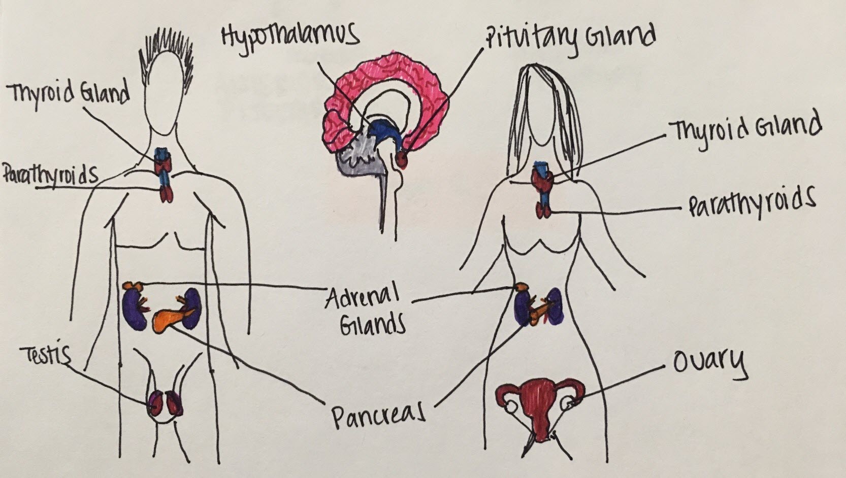
Figure 1. Main endocrine glands and their functions
Pediatrice Endocrine
A Chapter in Core Concepts of Pediatrics, 2nd Edition
Olivia Ginnard DO and Aikaterini Nella MD
Endocrinology is the scientific and medical discipline that focuses on hormones, including the function and disorders of endocrine glands.
The endocrine system consists of several glands that secrete hormones that affect the different organ systems (Figure1).
The main endocrine glands are the hypothalamus, pituitary gland, thyroid gland, parathyroid gland, adrenal glands, pancreas, and gonads (ovary and testis).
Figure 1. Main endocrine glands and their functions
Of the endocrine glands, the hypothalamus and pituitary glands are of major importance since they act as the coordinating centers of the endocrine system.
The hypothalamus is responsible for maintaining the body's internal balance (homeostasis) by stimulating or inhibiting major bodily functions such as the heart rate and blood pressure, body temperature, fluid and electrolyte balance, appetite and body weight, sleep cycle and function of the gastrointestinal track. The hypothalamus is also considered the master regulator of the endocrine system; Regulatory hormones secreted by the hypothalamus are transported by the hypophyseal-portal system to the anterior and posterior pituitary (Figure 2), prompting the release of secondary hormones that can affect various organ functions.
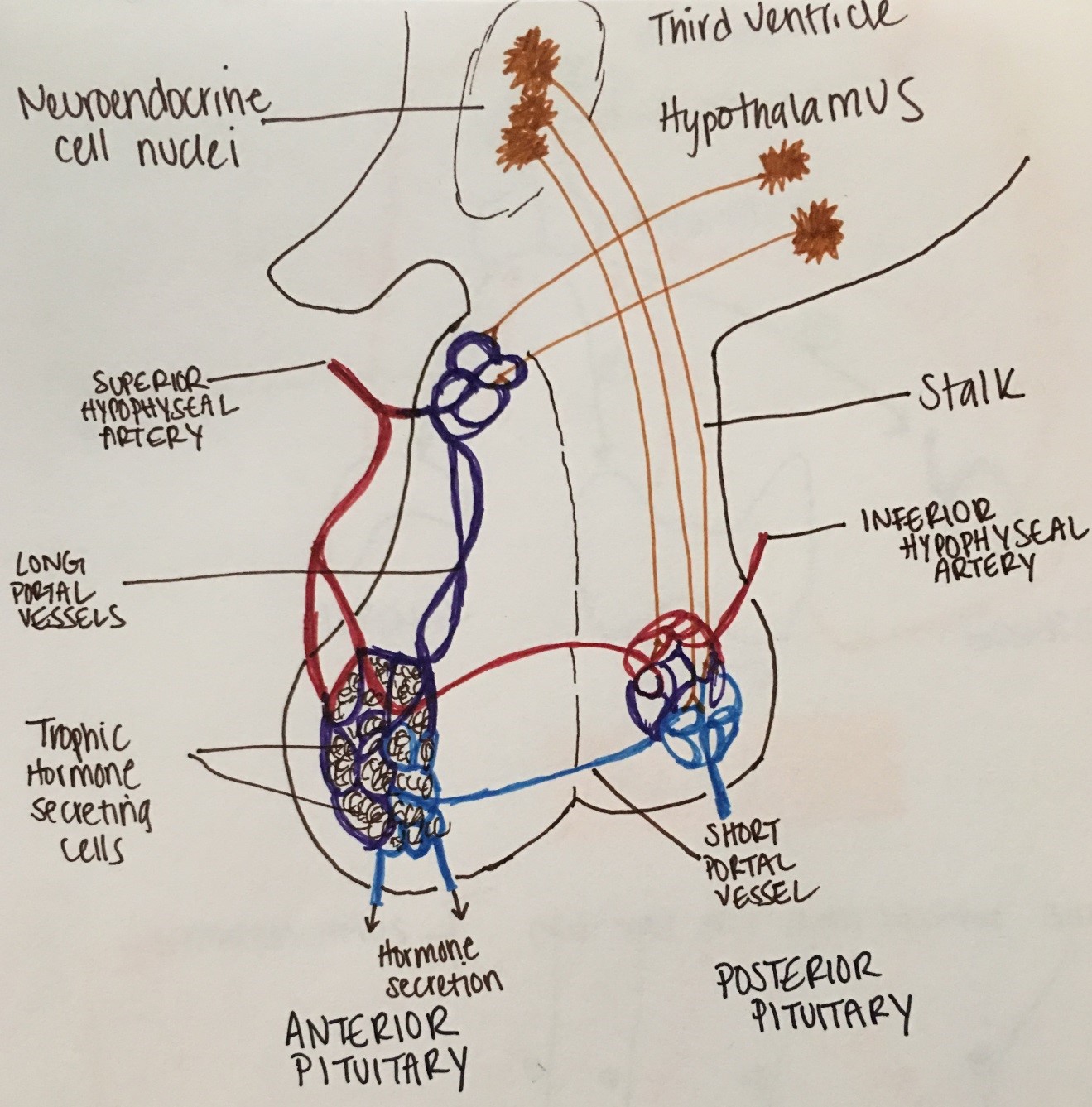
Figure 2. The hypophyseal-portal system
Hormones typically bind to membrane or nuclear receptors of specific target glands/organs, which in turn release hormones that exert negative feedback control on the releasing sites. (Figure 3).
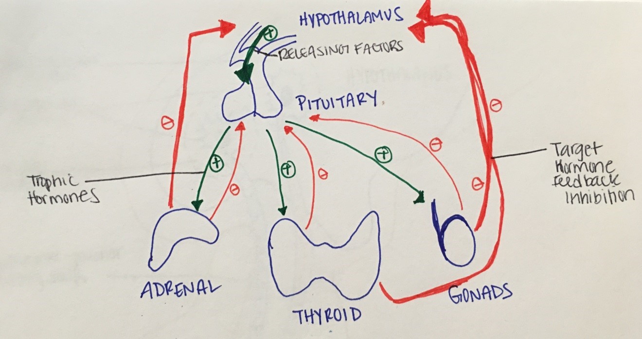
Figure 3. Example of negative (in red) feedback inhibition:
The hypothalamus secretes releasing factors that act on the pituitary gland to stimulate the release of trophic hormones.
Trophic hormones act then on target organs (e.g., adrenal, thyroid or gonads), which in response produce other
hormones/signals, shutting down the production of releasing or/and trophic hormones.
The hypothalamus secretes various hormones that are then transported to the anterior pituitary (Figure 4):
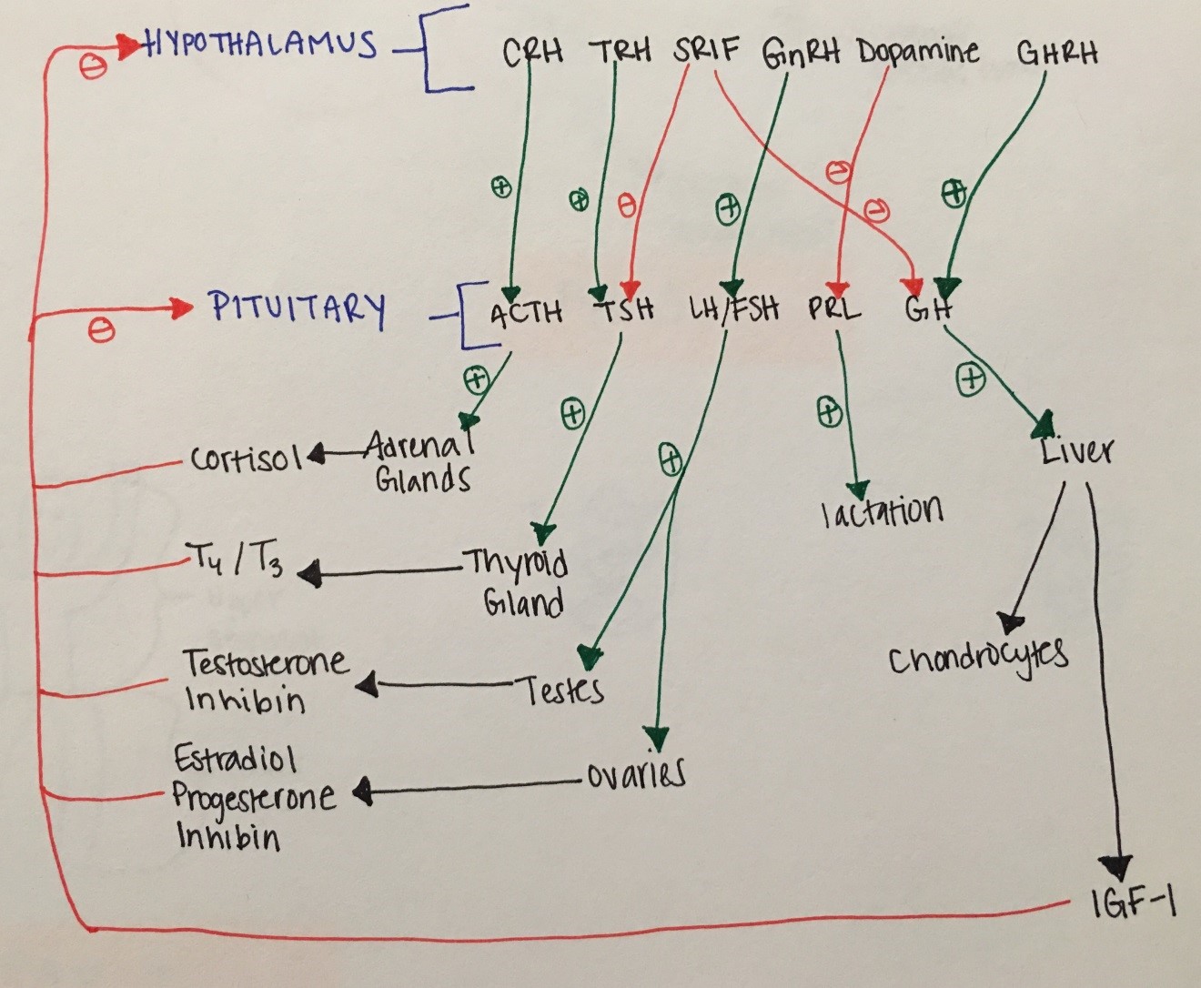
Figure 4. Overview of hypothalamic and pituitary hormones and their actions
The pituitary gland also secretes hormones in response to hypothalamic release hormones:
Supraoptic and paraventricular nuclei of the hypothalamus also secrete hormones that are transported to the posterior pituitary to be released into the circulation. These are:
Disorders of the hypothalamus can result in appetite, temperature and sleep disorders. As an example, hypothalamic obesity occasionally develops in response to major hypothalamic injury/damage affecting the centers of appetite regulation and energy balance. Hypothalamic obesity is characterized by an uninhibited eating disorder that often results in morbid obesity and can be associated with other obesity complications like diabetes, dyslipidemia, obstructive sleep apnea, mood disorder etc.
Disorders of the hypothalamus and/or anterior pituitary can also result in hypopituitarism, including adrenal insufficiency (see adrenal disorders section), hypothyroidism (see thyroid disorders section), hypogonadism (see puberty and its disorders section), growth hormone deficiency (see growth disorders section) and prolactin deficiency (inability to lactate).
Disorders of the posterior pituitary can result in diabetes insipidus and disorders pertaining to oxytocin deficiency (like inability to lactate, vaginal dryness, decreased libido etc.).
The neurohypophysis, or posterior pituitary gland, secretes vasopressin (AVP), also known as anti-diuretic hormone (ADH). AVP is synthesized by the supraoptic and paraventricular nuclei of the hypothalamus (see picture below), in response to plasma osmolality, intravascular blood volume changes (like bleeding, third spacing, etc.) and non-osmotic stimuli (e.g., nausea, drugs).
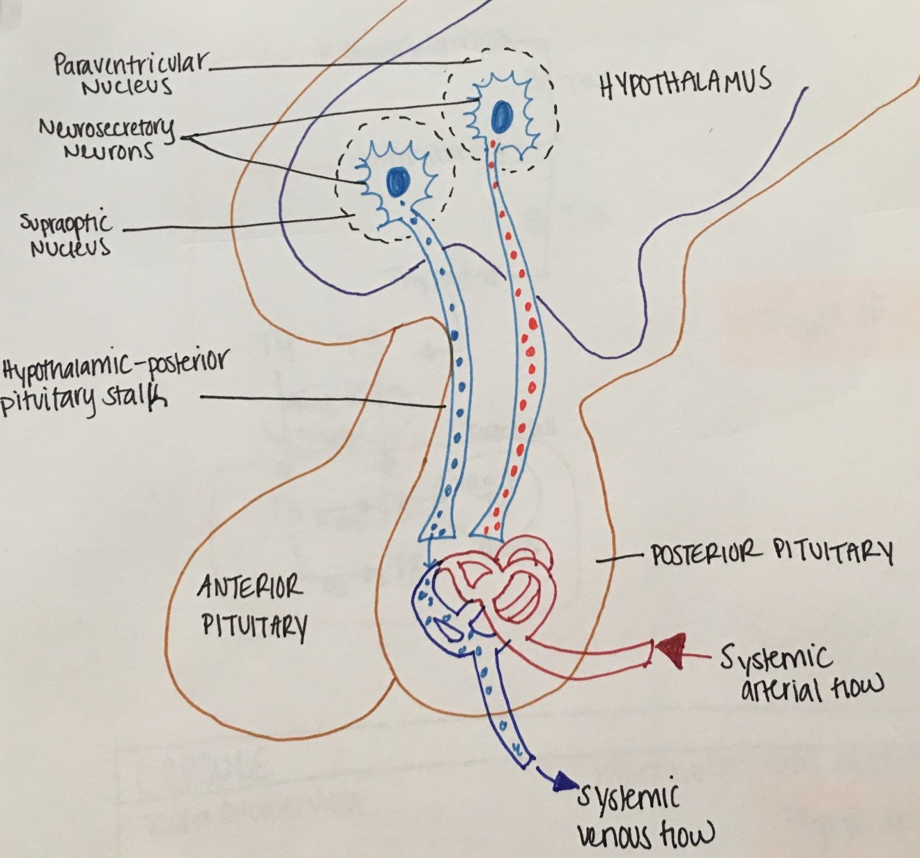
Figure 5. Physiology of the Hypothalamus and Posterior Pituitary
The antidiuretic effect of ADH is regulated through V2, cAMP dependent- receptors and aquaporing-2 proteins inducing increased water permeability and increased urea movement on the collecting ducts. In addition, ADH increases the rate of absorption of sodium (NaCl) in the thick ascending loop of Henle.
Diabetes insipidus (DI) is clinically defined as passing large volumes of hypotonic fluids (> 30ml/kg/24 hours with urine Osm < 300 mOsm/kg and specific gravity <1.010). DI can be central (due to deficiency of functional ADH) or nephrogenic (due to kidney inability to respond to the action of ADH). ADH may also be physiologically suppressed as a result of water overload (primary polydipsia). In contrast, in diabetes mellitus (DM), polyuria is due to osmotic diuresis (urine is hypertonic).
Etiology: Central DI or nephrogenic DI can be complete or partial, familial or acquired (e.g. brain tumor in central DI and lithium in nephrogenic DI).
Symptoms: polyuria and polydipsia, enuresis, FTT/poor growth, unexplained fevers, constipation
Diagnosis: elevated serum Na in patients with abnormal thirst mechanism and dilute urine (as above) vs failure to concentrate urine after a water deprivation test in patients with normal Na/ intact thirst mechanism
Treatment: monitor/maintain fluid balance (drink water as needed) and use ADH-agonists to prevent nocturia and minimize sleep disruption. ADH agonist include L-arginine Vasopressin (natural AVP-subcutaneous), DDAVP (synthetic- intranasal, IV or subcutaneous, or oral) and thiazide diuretics.
SIADH is due to excessive ADH secretion producing inappropriate urinary concentration and water retention, resulting in euvolemic hyponatremia.
Etiology: CNS disorders (head injury, CNS tumors, hydrocephalus etc.), pulmonary conditions (infections, cystic fibrosis), ectopic production (lung cancer etc.) drugs (thiazides etc.).
Symptoms: headaches, nausea, seizures, focal deficits
Diagnosis: low serum Na, low serum Osm, absence of signs of hypovolemia like tachycardia or hypervolemia like edema and ascites, absence of hypoadrenalism or hypothyroidism (that can also cause euvolemic hyponatremia)
Treatment: fluid restriction and drugs (e.g. vaptans- block ADH binding to V2 receptors)
Physical growth refers to bodily changes that happen as a child matures including weight, length or height and head circumference increases. Growth failure is by far one of the most common reasons for referrals to the pediatric endocrine office.
Linear growth is most rapid in prenatal life when it is mainly regulated by maternal and placental factors. Postnatal growth is progressively slower and predominantly reflective of childs own genetic potential (see growth velocity figure). Another growth acceleration occurs at puberty.
|
What is normal linear growth? |
|
|---|---|
|
0-1 years |
25 cm |
|
1-2 years |
10 cm/yr |
|
3-5 years |
7 cm/yr |
|
4-7 years |
6 cm/yr |
|
7 years - puberty |
5 cm/yr |
Table 1. Linea growth by age
Accurate measurement can not be overemphasized in the evaluation of the short child.
Growth Velocity: Growth velocity (cm/yr) = (Height2-Height1) / (#months between times) x 12. It determines normal or abnormal growth by comparing height change over a period of time to gender-appropriate norms.
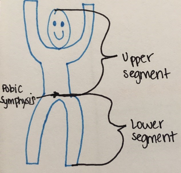
Figure 6. Measuring Upper to Lower segment ratio
Mid-parental height (MPH): measure of child's genetic height potential, using parents adult heights.
Mid parental height calculation:
Bone Age x-ray: tool for assessment of skeletal maturation as it compares to the chronological age and for the prediction of child's final adult height (for films older than age 6 years). Bone age x-ray is typically delayed in thyroid hormone and growth hormone deficiency or constitutional delay of growth, normal in familial short stature and advanced in precocious puberty.
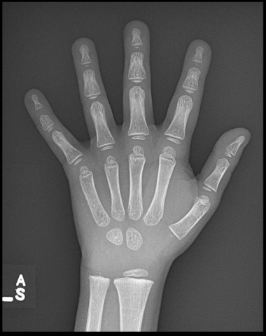
Figure 7. Example of bone age x-ray.
Children < the 3rd %ile on the growth chart or > 2 standard deviations below MPH deserve a short stature evaluation.
Common parental concerns about height tend to be gender biased (e.g. boys that are the shortest in their class or girls that are "too tall" or "taller than even her male classmates"). Referral for a short stature evaluation is also commonly prompted by concerns over bullying/ teasing at school for child's size/stature.
Diagnosis: Bone age x-ray (to assess skeletal maturation), CBC, CMP, ESR, UA to screen for systemic disease, free T4, TSH, IGF-1, and IGFBP-3 to screen for endocrinopathy. Celiac screen optional, Pituitary/brain MRI as needed. Karyotype/microarray or specialized genetic testing as clinically indicated.
Treatment:
Typically, growth hormone replacement is reserved for true growth hormone deficiency. Many causes of short stature are due to an underlying disease and by treating this disease, you are simultaneously able to treat the short stature. For example, levothyroxine treatment for hypothyroidism and growth hormone for growth hormone deficiency typically restores linear growth. However, growth hormone treatment is also FDA approved for Turner Syndrome, small-for-gestational age with failure to catch up, Prader-Willi Syndrome, idiopathic short stature, SHOX gene haploinsufficiency, Noonan Syndrome and chronic kidney disease.
Tall stature is defined as predicted adult stature greater than two standard deviations above the mean height for age and gender. Tall stature can represent a normal variant of growth such as familial tall stature or it may be pathological.
Etiologies:
|
Etiologies |
Example |
|---|---|
|
Genetic |
|
|
Acquired |
|
|
|
|
Syndromic Overgrowth |
|
|
|
Table 2. Common causes of pahological tall stature
Diagnosis: Bone age x-ray, thyroid function tests, fasting insulin level
Thyroid hormone is very important for the regulation of body metabolism and thermogenesis throughout life but it is of most critical importance for CNS development in the fetal and postnatal periods (especially the first 3 years of life). Thyroid hormone production is under the control of hypothalamic TRH and pituitary TSH but also iodine availability.
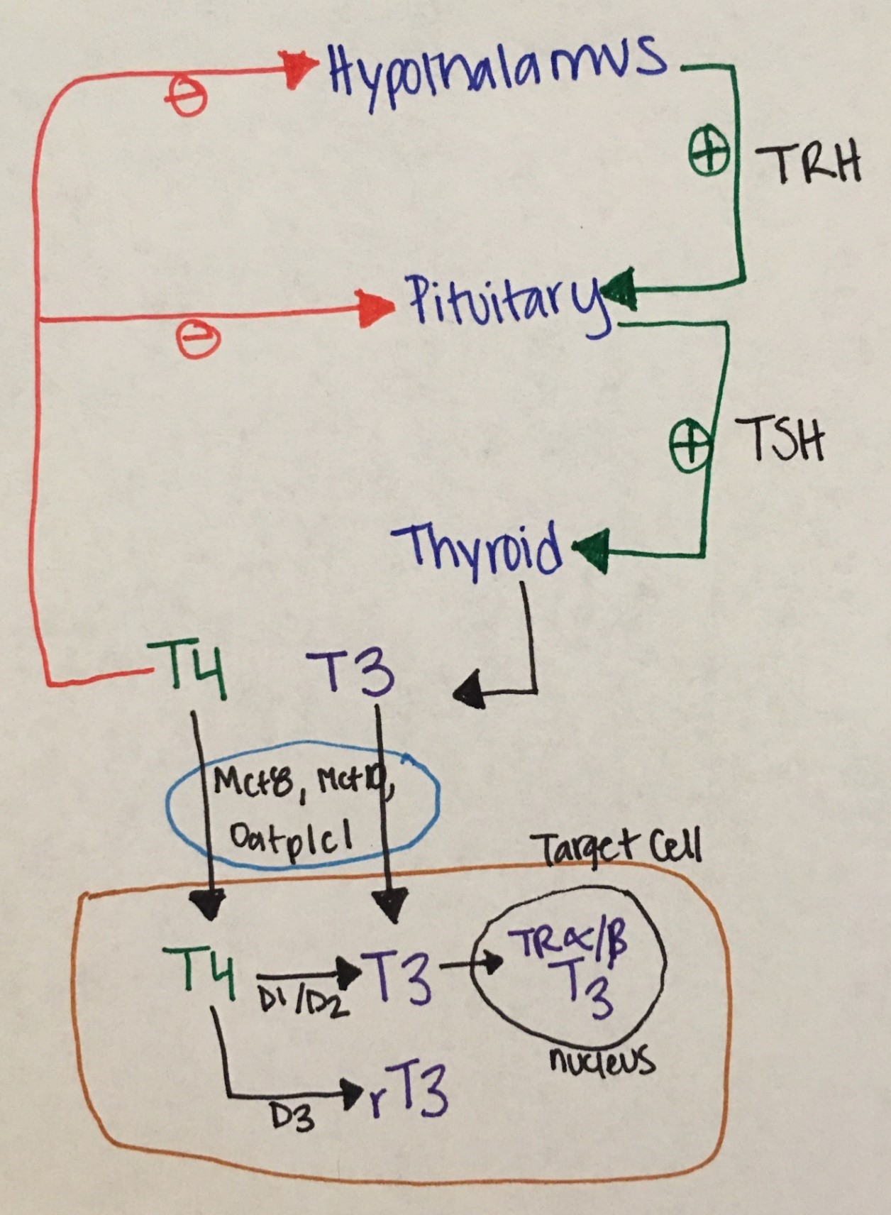
Figure 8. Phathophysiological Aspects of Thyroid Hormone Disorders/thyroid Peroxidase Autoantibodies and Reproduction
Fetal thyroid development: The fetal thyroid gland arises from an outpouching of the foregut and develops at the base of the tongue from where it will migrate down to its permanent location (over the thyroid cartilage) between the 4-8th weeks of gestation. It begins to concentrate iodine at about 10 weeks and hypothalamopituitary axis control is established at 18 weeks. Fetal T4 and TSH gradually rise to reach adult levels by 36 weeks of gestation. The rise in fetal serum T3 is less pronounced due to placental and fetal deiodeinases that covert T4 to the inactive hormone reverse T3 (rT3).
In response to the stress of birth and ambient temperature changes in the first 24 hours of life, TSH peaks up to 80 mIU/mL, driving T4 and T3 increases. Then all thyroid indices gradually normalize within the next several weeks (typically 4-6). Prematurity and perinatal complications may affect this normal process, resulting in delayed TSH rise or lower than expected T3/T4 levels.
Etiology: congenital vs acquired.
Congenital hypothyroidism (CH) is most commonly secondary to dysplasia/ hypoplasia of the thyroid gland or ectopia. In a minority of newborns, hypothyroidism is secondary to a defect in the synthesis of thyroid hormones (dyshormonogenesis), CNS/hypothalamic disorders affecting TRH or TSH synthesis, exposure to maternal antithyroid medication or as a result from transplacental transfer of TSH-receptor blocking antibodies. Outside the United States, the most common etiology of CH is iodine deficiency (or endemic goiter). Iodization of salt has significantly reduced CH in the United States.
Acquired hypothyroidism is most commonly due to Hashimoto thyroiditis, a destructive autoimmune disease in which there is lymphocytic infiltration of the thyroid gland. Other causes include post-surgical hypothyroidism (due to thyroid gland removal for hyperthyroidism or thyroid nodules/cancer), sick euthyroid syndrome, and subacute / suppurative thyroiditis. In autoimmune hypothyroidism (as also seen in autoimmune hyperthyroidism), children typically have a history or family history of autoimmune disease.
Symptoms:
Most newborns with CH appear normal at birth, unless mother was hypothyroid in which case babies may present with signs and symptoms of CH including jaundice (due to indirect hyperbilirubinemia), dry and coarse skin, umbilical hernia, constipation, thick protuberant tongue, poor muscle tone, anemia, hoarseness, growth retardation, delayed ossification and delayed closure of the anterior fontanel. Thyroid gland enlargement (goiter) is not usually present unless there is dyshormonogenesis. If babies with CH remain untreated, intellectual disability may ensue.
Children and teens with acquired hypothyroidism may present with decreased linear growth or even stunted growth, cold intolerance, constipation, dry skin, hair thinning or hair loss, sleepiness or irregular periods. A goiter and slow deep tendon reflexes may be present on exam. Delayed bone age maturation is also typically seen.
Diagnosis
Treatment includes levothyroxine. Dose and monitoring schedule is variable based on age of child. For newborns, dose is ~ 10-15 mcg/kg/day. Goal of treatment is to keep free T4 levels above the mean reference range with suppressed TSH.
Etiology. The most common etiology is Graves' disease (autoimmune stimulatory condition). Other rare causes include neonatal thyrotoxicosis due to maternal Graves' disease, activating TSH receptor mutation, and thyrotoxicosis as seen in the context of McCune-Albright Syndrome. Hot thyroid nodules are very rare in pediatrics.
Symptoms: include weight loss despite hyperphagia, palpitations, heat intolerance, increased perspiration, fatigue, diarrhea, hand tremor, anxiety and restlessness, goiter, rapid linear growth, irregular menses, and exophthalmos.
Diagnosis: TSH is typically suppressed (< 0.02 mIU/mL) and free T4, total T4 and T3 are elevated. Thyroid stimulating immunoglobulin is typically elevated.
Treatment: anti-thyroid drugs, radiation ablation (RAI) therapy or thyroidectomy. Beta- blockade is also typically necessary to decrease cardiac work.
Thyroid nodules are uncommon (incidence 1-1.5 %). Risk factors include female sex, pubertal age, medial or family history of thyroid disease. These typically present with an asymptomatic movable, soft and non-tender thyroid mass.
Thyroid cancer rare in children, typically presents with a painless or tender thyroid mass that is hard and fixated and associated with painless palpable neck lymphadenopathy. Distant metastases (neck, lungs, bones) are frequently present at the time of diagnosis, but survival/prognosis is excellent if early diagnosis and appropriate treatment are established. The most common type is papillary thyroid carcinoma (PTC). Follicular carcinoma is the second most common type of and is more prevalent in iodine-deficient regions. Medullary thyroid carcinomas are very rare and also the most aggressive, occurring most commonly as part of the familial MTC syndromes (e.g. MEN type 2A, MEN 2B, Carney complex etc.). The biggest risk factor is radiation exposure outside the familial cases.
The adrenal gland is divided into twodistinct areas, the cortex and the medulla. The cortex is further divided in 3 zones (zona glomerulosa or site of aldosterone synthesis, zona fasciculata or site of cortisol synthesis and zona reticularis or site of androgen synthesis). The medulla serves as the site of catecholamine, metanephrine and normetanephrine synthesis.
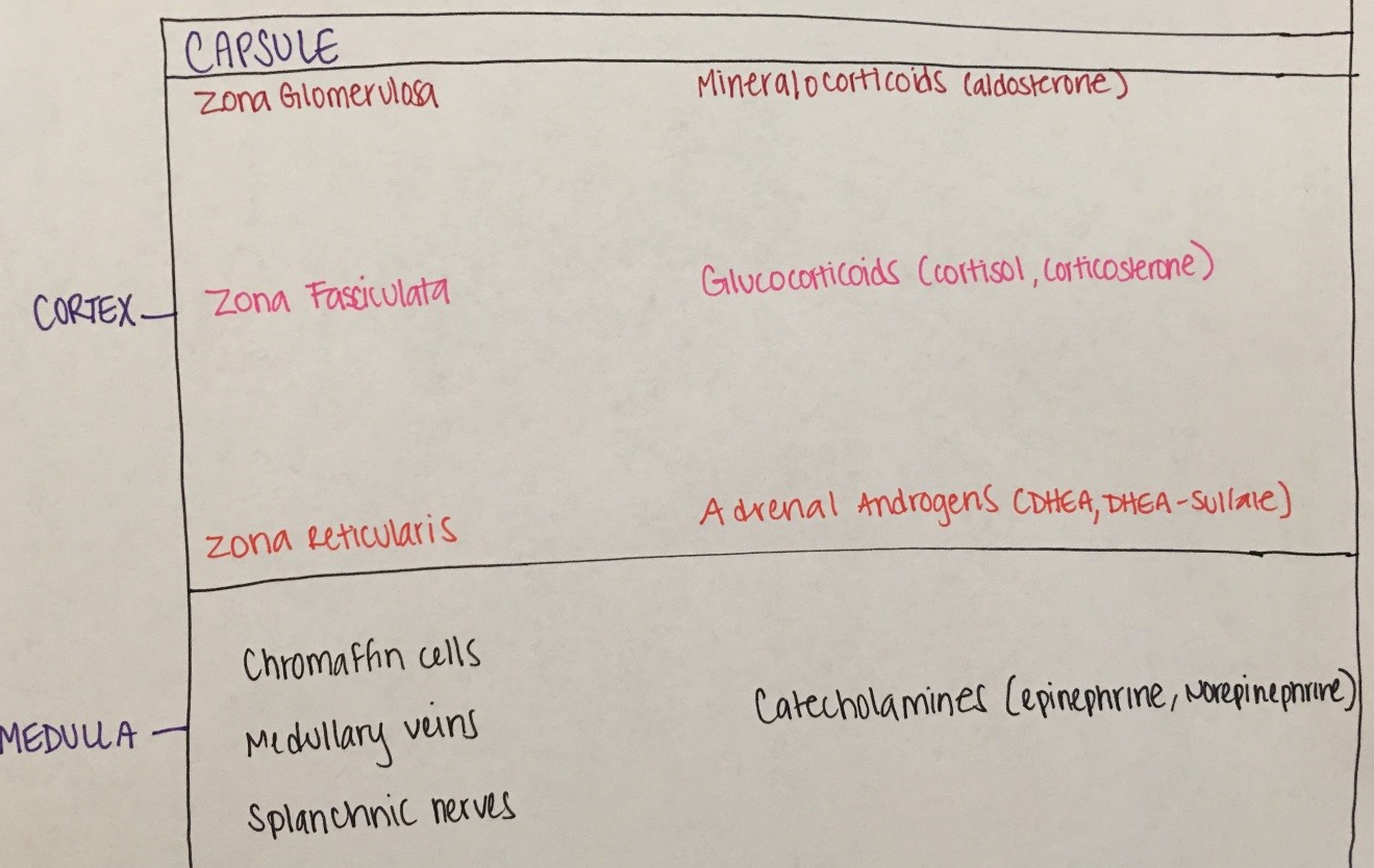
Figure 9. Adrenal glandareas and zones with respective hormones synthesized
Cholesterol (low-density lipoprotein or LDL) is the precursor for adrenal steroidogenesis. Adrenal steroidogenesis includes three main pathways, leading to cortisol, aldosterone and androgen synthesis, with androgens eventually being aromatized (converted to estrogens). See figure 10 below, where arrows represent enzymes that convert one substrate to another.
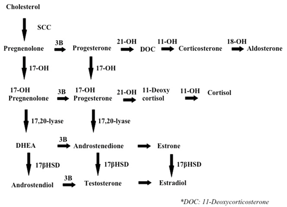
Figure 10: Main pathways of adrenal steriodogenesis
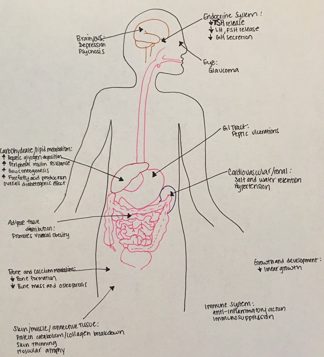
Figure 11: Multiple systemic effects of cortisol
Etiology: most commonly exogenous (iatrogenic or due to supraphysiologic exposure to glucocorticoids); rarely endogenous. Endogenous is most commonly due to excess pituitary ACTH secretion (Cushing disease or ACTH producing pituitary adenoma) and rarely due to ectopic ACTH production (e.g. lung or thymic tumor) or excess cortisol secretion by an adrenal tumor.
Symptoms: growth stunting, central obesity, facial plethora, buffalo hump, sleep disorders, hypertension, bruising, tinea corporis, and wide striae
Diagnosis: confirm hypercortisolism with low dose dexamethasone test vs midnight salivary cortisol vs 24-hr urinary free cortisol x3 days, if high cortisol check serum ACTH and refer to endocrinology for more specialized testing and imaging
Treatment: surgical removal of tumor, supplementary radiotherapy for Cushing disease
Etiology: most commonly iatrogenic (treatment with suprpaphysiologic doses of glucocorticoids can result to adrenal suppression) vs primary (adrenal gland is malfunctioning) vs central (pituitary or and hypothalamus are malfunctioning). Primary adrenal insufficiency can be autoimmune, due to congenital adrenal hyperplasia, adrenoleukodystrophy, adrenal hemorrhage etc. Central is due to isolated ACTH deficiency or associated with multiple pituitary deficiencies.
Symptoms: hypoglycemia, nausea, emesis, fatigue, anorexia, hypotension, hyponatremia, salt craving and hyperkalemia (if aldosterone deficiency), decreased pubic and axillary hair (if adrenal androgen deficiency), hyperpigmentation (due to high MSH).
Diagnosis: hyponatremia and hyperkalemia, high PRA, low aldosterone, elevated ACTH, failed ACTH stimulation test
Treatment: acute phase (IVF, IV hydrocortisone), chronic phase (glucocorticoids and fludrocortisones)
(discussed in detail under the ambiguous genitalia section)
Etiology: Catecholamine-secreting tumors, arising from chromaffin cells of the adrenal medulla. Very rare in children and usually inherited (MEN 2b, MEN 2a, NF-1, VHL, familial pheo/para etc).
Symptoms: flushing, sweating, palpitations, hypertension, headaches, emesis, and feeling of impending death
Diagnosis: Plasma or urine fractionated catecholamines/meta- and normetanephrines, CT adrenals and PET-FDG scan
Treatment: Removal of tumor with pre-op preparation to include alpha and beta blockade
Sex determination and sex differentiation can be divided into a) chromosomal sex or karyotype (46 XX, 46 XY, or variants), b) gonadal sex (presence of a testis or ovary) and c) phenotypic sex (external genitalia and internal structures).
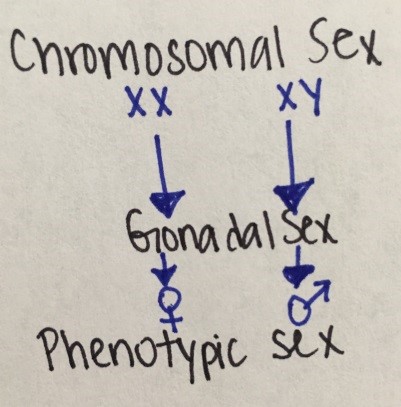
Figure 12. Sexual development
Human embryo is bipotential with genetic sex determined at conception. Male differentiation requires specific molecular and biochemical stimuli at specific times. Female differentiation occurs in absence of male determinant genes.
Disorders of sexual development can be associated with ambiguous genitalia (i.e., external genitals neither appearing clearly male nor female).
|
Sex Chromosome DSD |
46, XY DSD |
46, XX DSD |
|---|---|---|
|
45, X (Turner Syndrome & variants) |
Disorders of testicular development |
Disorders of ovarian development |
|
47, XXY (Klinefelter Syndrome & variants) |
Disorders in androgen synthesis or action |
Androgen excess |
|
45, X/46, XY (mixed gonadal dysgenesis) |
Other (i.e. hypospadias, congenital HH, cryptorchidism) |
Other (i.e.vaginal or uterine atresia) |
|
46, XX/46, XY (chimerism) |
|
|
Table 3. DSD classification table
The most common DSDs are:
Etiology: 47, XXY (classic) vs 46, XY/47, XXY (mosaic)
Symptoms: Tall stature, gynecomastia, small testes, azoospermia, learning difficulties
Diagnosis: High FSH and LH, low AMH and inhibin B, testosterone low in 50-75% of cases
Treatment: Testosterone replacement, spontaneous fertility unlikely
Etiology: 45 X (classic) vs Mosaic types (or mixed gonadal dysgenesis) like 45, XO/46, XX; 45, XO/46, XY etc.
Symptoms: Lymphedema, webbed neck/low hairline, high-arched palate, shield chest/wide-spaced nipples, short stature, streak ovaries, coarctation of aorta or other CV defects, learning difficulties
Diagnosis: prenatal typically. Based on cardiac anomalies, short stature in childhood, delayed puberty (high FSH & LH) in puberty.
Treatment: Estrogen/progesterone replacement therapy, long-term follow-up (cardiovascular, bone, reproductive health, hearing), spontaneous fertility unlikely, gonadectomy only when Y fragment contains TSPY locus (risk of gonadoblastoma increased)
Etiology: Group of autosomal recessive disorders, most common type is due to CYP21A2 gene mutations resulting in 21-hydroxylase deficiency. Classic (1:15,000 births; impaired cortisol synthesis). Non-classic (1:100- 1:1,000; cortisol synthesis relatively normal). Less common types include 11-beta hydroxylase deficiency, 17-hydroxylase deficiency, 3-beta- hydroxysteroid dehydrogenase deficiency.
Symptoms: in 21-hydroxylase deficiency, cortisol and aldosterone are not produced therefore patient present with adrenal salt/salt wasting symptoms. Also due to excess ACTH, excessive androgens are produced that can cause virilization of female infants or milder symptoms of hyperandrogenemia (like acne, premature pubarche, irregular periods) in the non-classic form. In 11-beta hydroxylase and 17-hydroxylase deficiency there is HTN due to excess mineralocorticoid and in 3-beta-hds there is adrenal insufficiency and androgen insufficiency, therefore males are under masculinized.
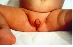
Figure 13 - Female infant with ambiguous genitalia (clitoromegaly, bifid scrotum, rugated folds) due to classic 21-hydroxylase deficiency.
Diagnosis: In 21-hydroxylase deficiency, newborn screen (NBS) detects most cases. If no NBS in place, classic type can present with hyponatremia, hyperkalemia, low aldosterone, high PRA, high 17-hydroxy progesterone, high androstenedione. With the non-classic type there is typically high serum 17-hydroxy progesterone, androstenedione and testosterone.
Management: For classic 21-hydroxylase deficiency: glucocorticoids, mineralocorticoids and also salt for infants. For non-classic 21-hydroxylase deficiency, glucorticoids may be needed but not always (depends on degree of hyperandrogenemia symptoms).
Most common cause of male undermasculinization
Etiology: 46, XY/ X-linked recessive/ Androgen Receptor (AR) gene mutation
Symptoms: Wide phenotypic spectrum:
Diagnosis: age-appropriate testosterone levels, elevated LH & FSH, normally formed testes, absent/vestigial Mullerian structures, variable presence of Wolffian ducts
Treatment: hormone replacement therapy +/- gonadectomy
Cryptorchidism mean that the testis is not in the scrotum and is not descended by 4 months old. This is the most common congenital abnormality in boys, present in 2-4% of term male infants. Complications include inguinal hernia, testicular torsion or trauma, subfertility, malignant transformation. Management involves orchidopexy (surgically moving the testes into the scrotum) or removal of dysgenic testis. Hormonal therapy (hCG/GnRH) may help testicular descent.
Normal female puberty. The hypothalamus-pituitary-ovarian (HPO) axis gradually increases in activity from the prepubertal age causing increases in LH, FSH, estradiol, inhibit A, and Inhibin B. These changes will eventually result in ovarian estrogen production and as a result breast growth, nipple and areola development, female body habitus, cornification of the vaginal epithelium, and enlargement of the uterus.
Normal male puberty. The increasing rise of LH and FSH causes an increase in testosterone, estrone, estradiol, androstenedione, 17-hydroxyprogesterone, Inhibin A, and DHEA. LH and FSH act directly on the testes to stimulated Leydig and Sertoli cell proliferation. Testicular testosterone causes enlargement of the penis, broadening of the shoulders with narrowing of the hips, deepening of the voice, beard growth, temporal hair recession, and thick/curly truncal hair.
Assessment: Tanner staging (or sexual maturation staging) by inspection and palpation is used to assess the sexual maturity of the child.
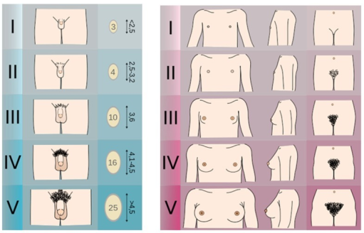
Figure 14. Tanner Stages
from https://openi.nlm.nih.gov/detailedresult.php?img=PMC4478390_fnhum-09-00344-g001&req=4
Definitions:
The onset of puberty in girls is defined by thelarche (breast budding) and it is typically between 8 and 12 years of age, although it can be as early as age 6 years in African American females. Puberty onset in boys is defined by gonadarche (testicular enlargement) and it is typically between 9 to 14 years of age. This is based upon data of predominately Caucasian children, but recent statistics reveal that other ethnicities may have earlier onsets of puberty. *
|
* Reference: AAP and Pediatric Endocrine Society, Precocious Puberty: A Guide for Families. 2012. https://www.aap.org/en-us/Documents/soen_precocious_puberty.pdf |
Adrenarche: increased adrenal androgen production measured by DHEAS. It precedes gonadarche and pubarche.
Gonadarche: increased activity of the HPO axis
Pubarche: onset of sexual hair growth
Thelarche: onset of breast development
Menarche: onset of first menstrual period
Precocious puberty is when the physical changes of puberty (thelarche with other secondary sex characteristics for girls, and gonadarche for boys) begin before the lower age limit for that sex, which is < 8 years for girls and < 9 years for boys. The lower limit is lower for other ethnicities such as African American or Mexican-American. Causes of precocious puberty can be gonadotropin dependent or gonadotropin independent.
Incomplete precocious puberty is isolated premature thelarche in girls < 3 years of age, isolated premature adrenarche, or isolated pubarche with slightly advanced bone age.
Etiologies:
|
GnRH-Dependent or Central |
Examples |
|---|---|
|
Idiopathic |
Sporadic or Familial |
|
CNS Abnormalities |
Acquired, Congenital |
|
Tumors |
|
|
Secondary to chronic exposure to sex steroids |
|
|
Reversible forms |
Space occupying or pressure associated lesions |
|
GnRH-Independent or Peripheral |
|
|
Genetic Disorder |
CAH, Gonadotropin-independent puberty, McCune-Albright |
|
Tumors |
Adrenal sex steroid secreting, Gonadotropin-producing, Ovarian, Testicular |
|
Limited or reversible forms |
Chronic primary hypothyroidism, CAH, Exogenous sex steroid or gonadotropins, Ovarian cysts |
Table 4: Etiologies of Precocious Puberty
Diagnosis: Evaluation includes suggestive physical exam findings, imaging in the form of bone age x-ray (typically advanced for chronological age), MRI of the head, pelvic/testicular ultrasound, and labs including LH, FSH, estradiol, testosterone, DHEAS, Free T4, TSH, and a GnRH stimulation test.
Treatment: includes treating the underlying cause, and if the cause is gonadotropin-dependent, then GnRH agonist therapy may be appropriate.
Delayed puberty is defined as lack of testicular enlargement by age 14 in boys and lack of thelarche by 13 in girls, no menarche by age 16, or no pubertal progression for 3 years after the onset of puberty. Delayed puberty can be due to central etiology (hypogonadotropic hypogonadism) or peripheral etiology (hypergonadotropic hypogonadism). Delayed puberty is most commonly due to "functional" hypogonadotropic hypogonadism such as in constitutional delay of growth and puberty, chronic illness, anorexia nervosa, etc. Pathological causes of delayed puberty can be divided into categories of primary hypothalamic-pituitary dysfunction and primary gonadal disorder.
Etiologies:
|
Etiologies of Pubertal Delay |
Examples |
|---|---|
|
Constitutional |
|
|
Hypergonadotropic hypogonadism |
|
|
Hypogonadotropic hypogonadism |
|
Table 5. Etiologies of Pubertal Delay
Diagnosis: Evaluation includes a physical examination, bone age XR, FSH/LH/testosterone/estradiol levels, cranial MRI, pelvic ultrasound, and karyotyping or genetic studies.
Treatment: short course of IM testosterone to trigger ("jump start") puberty in boys, oral estrogen/progesterone for puberty induction in girls. Long-term hormone replacement therapy may be needed in both sexes based on the nature of the problem and possibly continued androgen replacement therapy. Treatment for girls includes cyclic estrogen and progesterone.
Calcium, phosphorus, vitamin D, and the hormones that regulate them are very important in skeletal development and mineralization. Abnormalities in these can disrupt calcium homeostasis and therefore cause disorders of mineral metabolism or skeletal disorders of childhood.
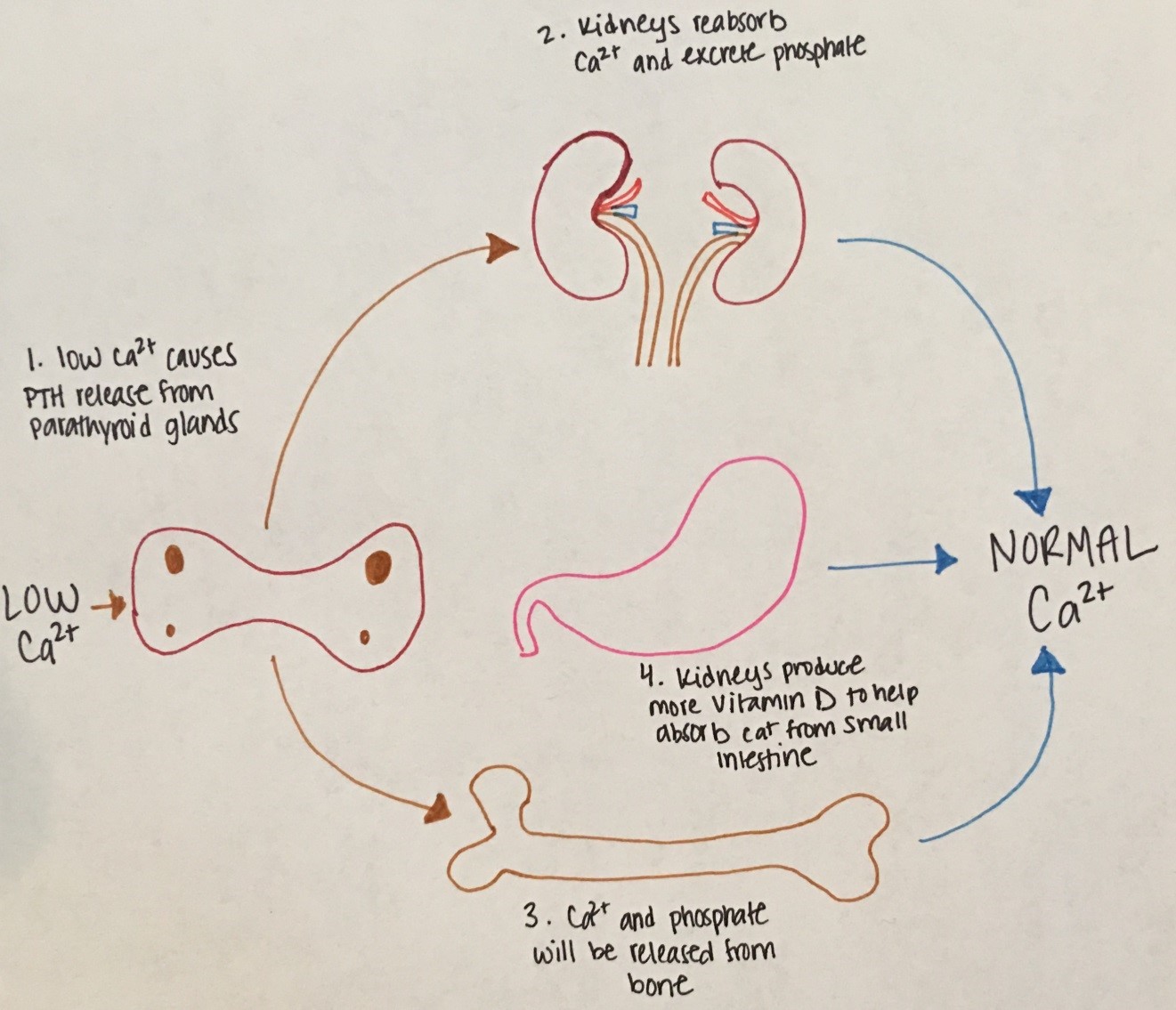
Figure 15. Low serum calcium stimulates release of PTH from the parathyroid glands. PTH stimulates calcium release
from bone and the kidneys to reabsorb calcium. The kidneys also excrete phosphorus and produce more vitamin D.
Vitamin D stimulates intestinal absorption of calcium.
Etiologies:
|
Types |
Physiology |
Examples |
|---|---|---|
|
Neonatal Hypocalcemia |
|
|
|
Illness |
|
|
|
Hypoparathyroidism |
|
|
Table 6. Etiologies of Hypocalcemia
Diagnosis: Measure serum calcium and vitamin-25OHD with corresponding phosphorus and PTH. Also, magnesium and alkaline phosphatase should also be checked. An ECG should be obtained to evaluate for changes related to hypocalcemia (i.e. prolonged QT interval).
Treatment:
Etiologies:
|
Types |
Physiology |
Examples |
|---|---|---|
|
Familial hypocalciuric hypercalcemia |
Hypercalcemia with elevated PTH, normal phosphorus, normal magnesium, elevated alkaline phosphatase, and low urinary calcium |
Mutation of CaSR gene |
|
Williams Syndrome |
Elevated calcium and calcitriol |
Facial anomalies, cardiac and renal defects, hyperacusis, and cognitive impairment |
|
Hyperparathyroidism |
Elevated calcium, elevated PTH, low phosphorus, and elevated calcitriol |
Childhood neck irradiation |
|
Immobilization |
Decreased skeletal mineral Elevated serum calcium and urinary calcium |
Prolonged immobilization Weightless space travel |
|
Malignancy |
Osteoclastic activity leading to excessive calcium release from bone |
Lymphomas Squamous cell carcinoma Breast cancer Renal cell carcinom, bladder carcinoma Multiple myeloma |
Table 7. Etiologies of Hypercalcemia
Diagnosis: Measure calcium, phosphorus, PTH, magnesium, Vitamin-25OHD, and alkaline phosphatase. ECG will show shortened ST segment and heart block.
Treatment: Increase urinary excretion of sodium with furosemide. Start calcitonin to indirectly inhibit osteoclastic bone resoprtion or inhibit bone resorption directly with bisphosphonates.
Rickets occurs when there is defective mineralization of the epiphyseal growth plate. Although there are different causes of rickets, all forms show characteristic widening and flaring of the epiphyses.
Etiologies:
|
Causes |
Examples |
|---|---|
|
Vitamin D deficiency |
|
|
Calcium Deficiency Rickets |
Unclear underlying causes |
|
Rickets of prematurity |
|
|
Hypophosphatemic Rickets |
|
Table 8. Etiologies of Metabolic Bone Disease.
Glucose is an important source for brain energy metabolism and extensive regulatory mechanisms are in place to ensure protection from hypoglycemia. Glucose concentrations naturally reach a nadir a couple hours after birth and then begin to rise reaching normal values by day 3 of life. This is related to the abrupt cessation of placental glucose transfer at delivery causing a transient decrease in glucose levels with subsequent response of increased glucagon, decreased insulin levels, and an increase in catecholamine secretion to gradually normalize plasma glucose concentration. Regulatory mechanisms in older children are balanced by gluconeogenesis and glycogenolysis. Hypoglycemia definition varies based on the age group (40 mg/dL or below in neonates) and < 55-60 mg/dL in older children.
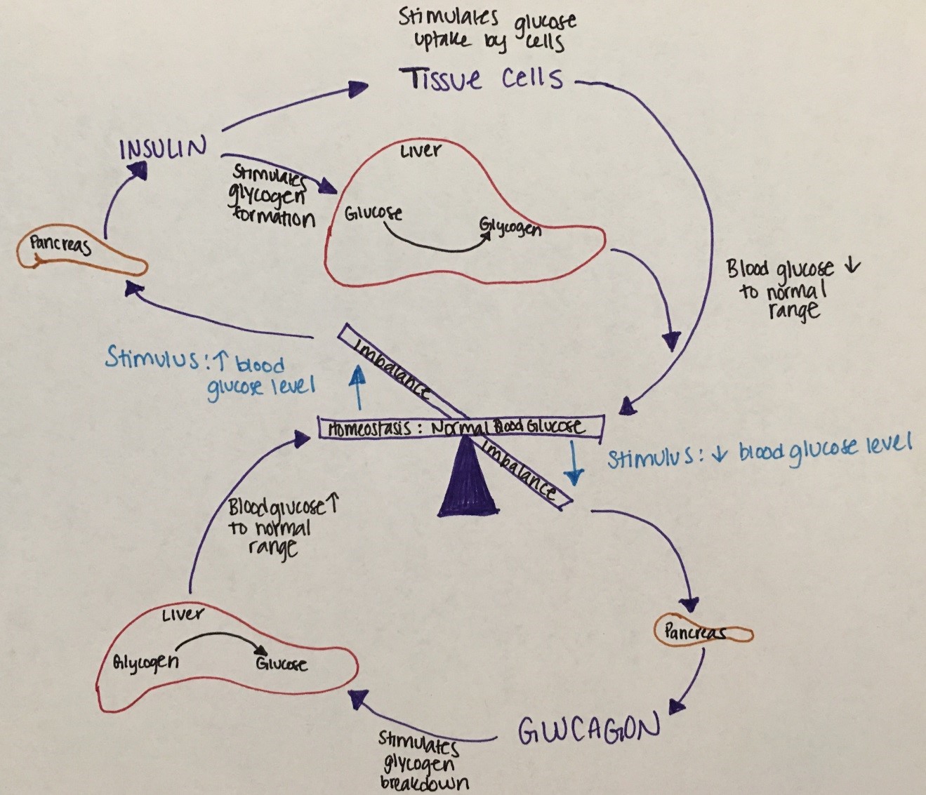
Figure 16. This figure describes the balance of glucose regulatory mechanisms.
Lower blood glucose levels stimulate the pancreas to release glucagon to stimulate glycogen breakdown in the liver to increase blood glucose levels.
High blood glucose levels stimulate insulin release from the pancreas to stimulate glycogen formation in the liver to lower blood glucose levels.
Etiology: Hypoglycemia is due to defects of the hormones or enzymes of the glucose regulatory mechanisms that result in inadequate glucose or surplus of insulin.
|
Age |
Etiologies |
|---|---|
|
Infant |
|
|
Toddler |
|
|
Child |
|
Table 9. Etiologies of Hypoglycemia by Age
Symptoms: An abrupt decrease in plasma glucose causes adrenergic symptoms (due to catecholamine release) such as pallor, sweating, tachycardia, tremor, and emesis. A slow decrease in plasma glucose causes neuroglycopenic symptoms such as confusion, diplopia, headache, dizziness, lethargy, seizure, and lack of coordination. Neonatal symptoms of hypoglycemia are more subtle, such as apnea, low temperature, poor tone, jitteriness, and poor feeding.
Diagnosis: Critical sample needs to be obtained when BG at or <40 mg/dL and includes serum free fatty acids, ketones, and insulin levels. Hormones to be also obtained include cortisol, ACTH, and growth hormone. Acylcarnitine and total carnitine, urine organic acids and serum amino acids to be considered based on the age group/ index of suspicion. If a child has hepatomegaly with hypoglycemia, suspect an enzyme deficiency. Males with a microphallus and hypoglycemia should be evaluated for hypopituitarism. Accidental ingestion of alcohol or salicylates can cause hypoglycemia so toxicology screen and history taking will help with diagnosis in this instance. Finally, if Munchausen by Proxy is suspected, then c-peptide along with insulin levels need to be obtained to investigate for exogenous insulin administration.
Treatment: Acute treatment includes early feeding (neonates), dextrose gel, or IV boluses of 2 mL/kg of 10% dextrose in water followed by continuous infusions of glucose. If hypoglycemia persists, it is then treated with continuous glucose infusion, diazoxide and/or somatostatin analogs. If hypoglycemia persists, pancreatectomy may be necessary.
Diabetes mellitus (DM) is a common metabolic condition of hyperglycemia caused by complete or partial insulin deficiency and its actions.
Insulin physiology: Insulin is made on the ribosomes of pancreatic islet beta cells as a proinsulin precursor (single chain: chain A is connected to chain B by c-peptide) that is then cleaved into insulin and c-peptide molecule. It is then released into circulation as a double chain polypeptide linked by disulfide bridges. C-peptide has a longer half-life and later nadir.
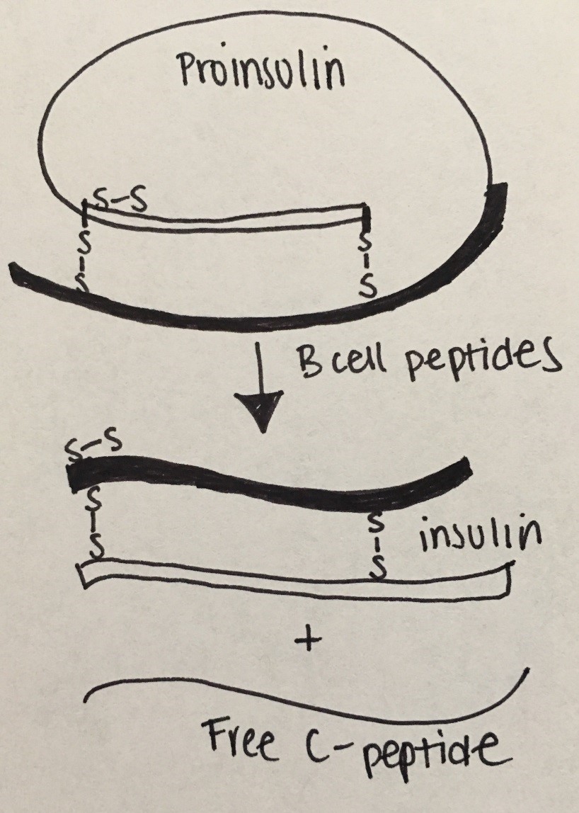
Figure 17: Insulin Physiology
Etiologies: There are many subgroups with distinct etiologies but the most commonly known will be discussed.
Type 1 DM (previously referred to as Juvenile Onset Diabetes): T1DM is due to autoimmune destruction of pancreatic beta cells resulting in insulin deficiency. Histocompatibility locus antigens that determine response to self or other antigens create antibodies to cytoplasmic and cell surface components of the islets cells, to insulin, to GAD (glutamic acid decarboxylase or enzyme in pancreatic islet innervation), and to IA-2 (islet phosphatase). T1DM is the most common type in childhood and adolescence (7-15 year olds); but with the epidemic increases in childhood obesity, there is an increased prevalence in T2DM among this age. Wasting and diabetic ketoacidosis are complications from undiagnosed/ untreated hyperglycemia.
Type 2 DM: T2DM is typically due to insulin resistance with some B-cell impairment. The insulin resistance can be observed at the level of skeletal muscle, liver, and adipose tissue, but can result in actual insulin deficiency. For more info regarding the differences between T1DM and T2DM see table below:
|
|
Type 1 |
Type 2 |
|---|---|---|
|
Insulin Deficiency: |
Absolute |
Relative |
|
Age of Onset |
75% < 21 yrs.
|
75% > 40 yrs |
|
Weight |
Thin |
Overweight |
|
Edtiology: |
Autoimmune |
Sedentary lifestyle |
|
Inheritance: |
HLA-Assn |
Complex |
|
Treatment: |
Insulin |
Healthy nutrition, excercise, oral agents |
Table 10. Comparison of Type 1 and Type 2 Diabetes Mellitus.
Monogenic diabetes (MODY or maturity onset of diabetes of youth). MODY has a dominant inheritance pattern with the mutations of the genes encoding enzymes or transcription factors affecting islet cell development and insulin secretion. This causes GLUT2 transporter regulation to be mutated.
Symptoms: polyuria, polydipsia, weight loss (T1DM); obesity (T2DM) and acanthosis nigricans. Complications: retinopathy, nephropathy, neuropathy, ischemic heart disease, vasculopathy, diabetic ketoacidosis, nonketotic hyperosmolar coma.
Diagnosis: Initial laboratory workup includes random BG or fasting BG, HbA1C, UA. BMP and blood gas are typically obtained if there is concern for DKA. Celiac panel, thyroid function tests, c-peptide and autoimmune diabetes antibodies studies (i.e. anti-GAD, anti-islet, etc) are obtained if T1DM is suspected.
Laboratory findings between T1DM and T2DM
|
|
T1DM |
T2DM |
|---|---|---|
|
Insulin |
Low |
Normal or Elevated |
|
Random Glucose |
> 200 |
>126 |
|
Background |
Autoimmune |
Genetic basis |
|
Ketosis |
Yes |
No |
|
Associated Diseases |
APS-1, Addison's Disease |
Metabolic Syndrome |
Table 11. Laboratory findings for T1DM and T2DM
|
|
Normal |
Mild |
Moderate |
Severe |
|---|---|---|---|---|
|
CO2 |
20-28 |
16-20 |
10-15 |
<10 |
|
pH |
7.35-7.45 |
7.25-7.35 |
7.15-7.25 |
<7.15 |
|
Clinical |
No change |
Oriented but fatigued |
Kussmaul respirations, oriented but sleepy |
Kussmual or depressed respirations; sleep to depressed sensorium or coma |
Table 12. Classification of DKA (from Nelson Textbook of Pediatrics)
Treatment:
T1DM: Patients are typically dependent on exogenous insulin; fluids; diet.
T2DM: First line treatment entails a diabetic diet (healthy, sugar avoidance, and CHO restriction) coupled with increased physical exercise. Oral hypoglycemic are typically introduced at the time of diagnosis. Exogenous insulin treatment is reserved for severe forms that have had DKA or HgA1C > 9%.
Obesity is defined as BMI > 95th percentile for age and sex. Overweight is defined as BMI of 85-95th percentile. Increasingly common in children (US prevalence 18.5% or ~ 13.7 million children and adolescents). Obesity is associated with risk for comorbidities that can persist into adulthood, such as diabetes mellitus, hypertension, early onset cardiovascular disease and osteoarthritis, dyslipidemia, precocious puberty, polycystic ovarian syndrome etc.
Etiology: most commonly exogenous (usually due to a combination of genetic predisposition and dietary/lifestyle habits). Other etiologies include drug-induced and endocrine-related. Genetic conditions associated with obesity include Prader-Willi Syndrome, Alstrom Syndrome, Bardet-Bied Syndrome, Down Syndrome, and Turner Syndrome. Endocrinopathies that can be associated with obesity include Cushing Syndrome and hypothalamic obesity and less likely growth hormone deficiency or hypothyroidism. Antidepressants, antiepileptics, glucocorticoids, and atypical antipsychotics are associated with an increased risk of obesity.
Diagnosis: Based on physical exam/ BMI. Screening for co-morbid conditions as sleep apnea, non-alcoholic fatty liver disease, type 2 diabetes, hypertension, dyslipidemia, or polycystic ovary syndrome is imperative.
Treatment: dietary/lifestyle changes to promote weight loss. The only FDA approved medication for obesity in children less than 16 years of age is Orlistat but has unfavorable side effects, such as flatulence and oily, loose stools. Bariatric surgery can be considered for children with BMI greater than 40 kg/m2 with near-complete skeletal maturity.
Lipids are organic and water insoluble compounds and include fatty acids, triglycerides, and cholesterol. Lipoproteins (LDL, IDL, VLDL, HDL, and chylomicrons) are soluble lipid or fat proteins that facilitate lipid movement within the aqueous environment of the body.
After digested fat is absorbed, chylomicrons are produced in the intestinal lumen and then transferred to adipose tissue, muscle, and the liver. There chylomicrons will be hydrolyzed to free fatty acids or VLDL that in turn will be metabolized to LDL (the major carrier of cholesterol to tissues). HDL acts as a "bad fat" scavenger, transferring excess cholesterol from peripheral tissues to cells that require it or to the liver to be excreted. Any abnormalities in these pathways or metabolism can lead to lipid disorders.
Etiology: The prevalence of dyslipidemia in children age 12-19 was reported as approximately 20.3% in the CDC Morbidity and Mortality Weekly Report. The most common etiology of dyslipidemia is exogenous (usually due to excessive simple carbohydrate and saturated fat intake with limited physical activity). Other etiologies include genetic (familial hypertriglyceridemia, familial hypercholesterolemia, familial defective apoB100, familial combined hyperlipidemia etc.), drug-induced (glucocorticoids, isotretinoin, protease inhibitors, immunosuppressives, beta blockers), and endocrinopathies (hypothyroidism, hyperthyroidism, T2DM, Cushing Syndrome).
Symptoms: usually associated with obesity and acanthosis nigricans. Hypertriglyceridemia may be associated with lipemia retinalis and cutaneous eruptive xanthomata. Hypercholesterolemia can be associated with xanthelasmas, corneal arcus, and tuberous xanthomata.
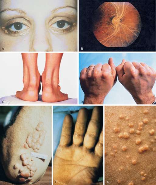
Figure 18 - Physical examination findings associated with hyperlipidemia.
From Williams Textbook of Endocrinology by Shlomo Melmed, pg. 1678.
A, Xanthelasma. B, Lipemia retinalis. C, Achilles tendon xanthomas. Notice the marked thickening of the tendons.
D, Tendon xanthomas. E, Tuberous xanthomas. F, Palmar xanthomas. G, Eruptive xanthomas.
Diagnosis: Universally screen children aged 9-11 years with a non-fasting or fasting lipid profile. A diagnosis of dyslipidemia is made with total cholesterol greater than 169 mg/dL, LDL greater than 109 mg/dL, non-HDL greater than 119 mg/dL, Apo-B greater than 89 mg/dL, triglycerides greater than 74 mg/dL in children aged 0-9 years or greater than 89 mg/dL in children older than 9 years old, or HDL less than 45 mg/dL
Treatment: diet (CHILD-1 and CHILD-2) and lifestyle changes is the mainstay. Statins are considered for cases of severe hypercholesterolemia or moderate hypercholesterolemia with risk factors/comorbidities if there is no response to a minimum of 6 months of diet and lifestyle intervention.
There are syndromes with a constellation of multiple endocrine diseases termed polyendocrinopathies.
|
|
APS-1 |
APS-2 |
IPEX |
|---|---|---|---|
|
Inheritance Pattern |
AR |
Polygenic |
X-linked |
|
Gene Associated |
AIRE |
HLA allele DR allele |
FOXP3 |
|
Age of Presentation |
Infancy 3-5 years old Early adolescence |
20-40 years old |
|
|
Associated Diseases |
Chronic mucocutaneous candidiasis Hypoparathyroidism Addison's Disease |
Addison's Disease Thyroid disease T1DM
|
Immune dysfunction Polyendocrinopathy Enteropathy Treatment and Screening: Treatment is primarily focused on the underlying diseases. Family members of patients with APS-2s should have thyroid function tests every 5 years due to the most commonly found endocrinopathy being hypothyroidism.
Multiple endocrine neoplasia (MEN1 and MEN2) |
Treatment and Screening: Treatment is primarily focused on the underlying diseases. Family members of patients with APS-2s should have thyroid function tests every 5 years due to the most commonly found endocrinopathy being hypothyroidism.
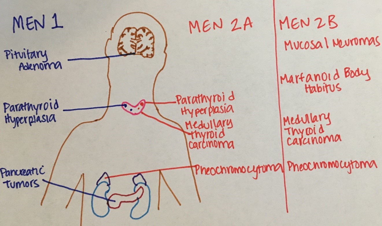
Figure 19. Comparison of MEN type
Etiology: MEN 1 follows an autosomal dominant inheritance pattern due to a mutation of the MENIN tumor suppressor gene. It is rare before the age of 10 years and typically peaks in the second and fourth decades of life. MEN2 also follows an autosomal dominant inheritance but associated with a RET gene mutation.
Symptoms and Diagnosis: Hyperparathyroidism is typically the presenting feature of MEN1. Suspected patients should have screening labs that include serum calcium, prolactin, gastrin, and pancreatic polypeptides. Signs that are consistent with hypercalcemia include lethargy, depression, constipation, kidney stones, and shortened QT interval on ECG. Symptoms of pituitary tumors include findings consistent with prolactinomas (galactorrhea or sexual dysfunction), acromegaly, hyperthyroidism, Cushing's disease, and visual disturbances. With MEN1, you may find high PTH, calcium, gastrin, insulin, and VIP on biochemical studies. MEN2 typically has high calcitonin related to medullary thyroid carcinoma and elevated plasma or urine catecholamines related pheochromocytoma. Imaging of the pancreas and pituitary should also be obtained.
Treatment: Primarily focused on treating the underlying diseases. Family members should be screened, as well, due to the inheritance pattern and high penetrance.
This syndrome is defined by a clinical triad of fibrous dysplasia of bone, café au lait spots, and precocious puberty. Growth hormone excess, hyperthyroidism, and Cushing Syndrome can also be seen. Associated with mutation of GNAS.
Treatment: Fibrous dysplasia can be followed clinically with annual imaging of the skull and mandible and annual hearing/vision testing. Sometimes, surgery or bisphosphonates may be warranted in severe cases. Precocious puberty is typically treated with aromatase inhibitors or estrogen agonists. Close monitoring is required for testicular lesions in males.
Unless otherwise noted:
All unspecified drawings were created by Olivia Ginnard MD University of Texas Medical Branch and used with her permission.