Arrhythmias
Arrhythmias are defined as disturbances in heart rate and/or conduction. Arrhythmias result from abnormal impulse formation, abnormal impulse conduction, or both. Arrhythmias may occur in children with normal hearts and/or may be associated with CHD, medications or electrolyte disturbances.
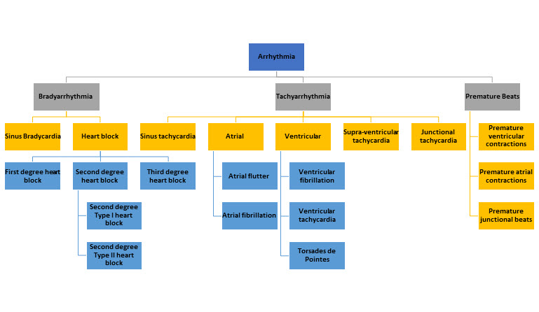
Types of Arrrhyhimias
Bradyarrhythmias
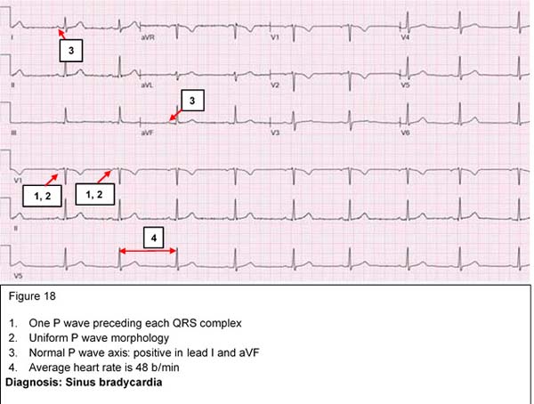
Sinus bradycardia
The normal range of heart rate depends on the age of the individual, ranging from 120-160 beat/min in the newborn to 60-80 beat/min in the adult. Trained athletes may normally have sinus bradycardia due to increased vagal tone. Pathological sinus bradycardia is usually secondary to an underlying condition such as hypothyroidism or medications such as beta-blockers.
Asymptomatic physiologic sinus bradycardia requires no treatment. In symptomatic bradycardia, the underlying cause should be treated and a pacemaker placement may be considered if there is no response to medical therapy.
Atrioventricular Block:defect of conduction in the AV node
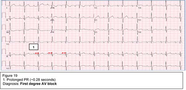
First degree AV block
This indicates prolongation of the PR interval more than 95th percentile for age and heart rate. At any age, PR interval > 0.2 seconds is considered prolonged.
Causes include:
- Impairment in the AV node conduction caused by increased vagal tone
- AV nodal ischemia
- Drugs such as digoxin and beta-blockers
It is usually reversible and does not require any treatment. First degree AV block could be one of the cardiac manifestations of rheumatic fever.
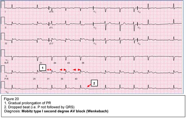
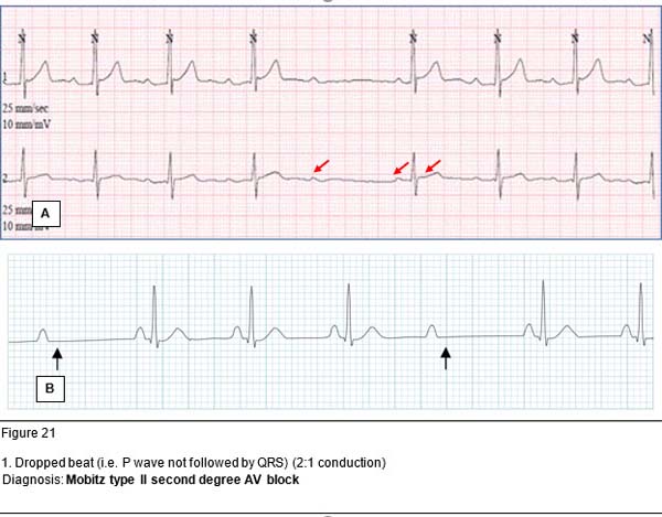
Second degree AV block
This is secondary to an intermittent failure of conduction through the AV node so that some P waves are not followed by QRS complexes.
Mobitz type I (Wenckebach) is a gradual prolongation of the PR interval until there is a P wave that is not conducted (not followed by a QRS complex). It is usually benign and may be seen in the presence of increased vagal tone, during sleep or in trained athletes.
Mobitz type II is sudden loss of AV conduction (two or more P waves before QRS complexes). It is more serious as it may progress to a complete AV block. Implantation of a pacemaker may be considered in symptomatic patients.
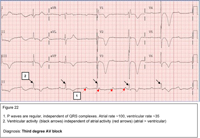
Third degree (complete) AV block (atrioventricular dissociation)
Complete AV block represents complete atrioventricular dissociation with no correlation between the atrial and ventricular electrical activity. The ventricular rate is significantly slower than the atrial rate. A pacemaker placement is warranted in symptomatic patients. This condition may be seen in infants born to mothers with systemic lupus erythematosus (SLE).
Tachyarrhythmias
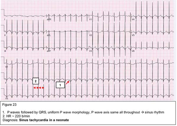
Sinus tachycardia:
Sinus tachycardia is characterized by narrow fast QRS complexes that are preceded by normal P waves. Sinus tachycardia may be a physiologic response to exercise, anxiety, fever, hypovolemia, hypoxemia or hyperthyroidism. A good rule of thumb to remember for fever is 1 degree Celsius increase in temperature accounts for ~10 b/min increase in HR. Maximum physiologic HR is 220 bpm age in years.
Maximum heart rate =220 - age in years
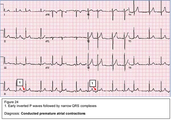
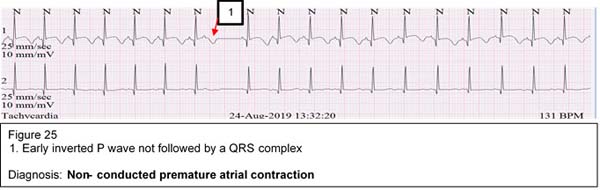
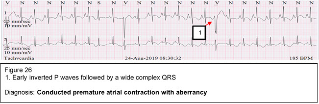
Premature atrial complexes (PAC's)
PAC's represent an early atrial electrical activity outside the SA node. PAC's may appear in one of three forms:
- Premature P wave followed by a narrow QRS complex (conducted PAC)
- Premature P wave not followed by a QRS complex (non-conducted PAC)
- Premature P wave followed by wide QRS complex (conducted PAC with aberrancy i.e. bundle branch block)
PAC's are commonly seen in infants and usually disappear with increasing age. It is usually benign and needs no treatment.
Supraventricular Tachycardia (SVT)
SVT is characterized by a narrow QRS complex tachycardia with a heart rate of 250-350 beat/min. It is commonly seen in children with normal hearts but may be associated with some CHD such as Ebstein anomaly. SVT may be caused by an accessory pathway between the atria and the ventricles or by a reentry circuit within the AV node. In infants, SVT presents with poor feeding, irritability, sweating and respiratory distress. Prolonged SVT may lead to CHF due to coronary insufficiency.
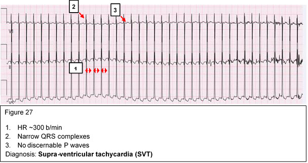
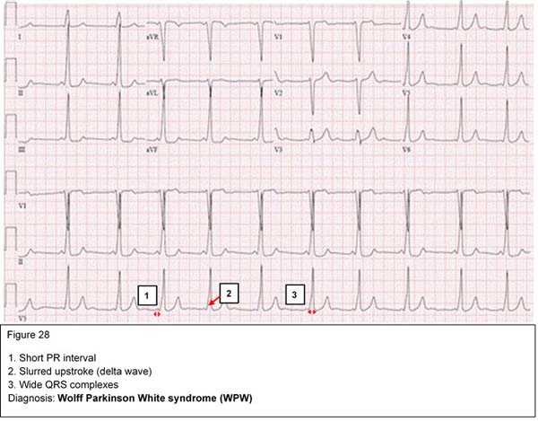
Compensated SVT should be treated promptly with vagal maneuvers such as application of ice to the face. If this is unsuccessful, then adenosine should be administered intravenously. Children with uncompensated SVT should undergo cardioversion. Wolff Parkinson White (WPW) syndrome is an example of pre-excitation due to an accessory pathway between the atria and ventricles. It is characterized by short PR intervals, delta waves, and wide QRS complexes.
Ventricular arrhythmias
Ventricular arrhythmias are characterized by wide QRS complexes that are not preceded by P waves, and abnormal T waves. The symptoms depend on the heart rate and are usually due to poor ventricular filling. This is a potentially serious dysrhythmia and synchronized cardioversion is commonly indicated.
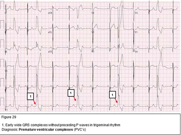
Premature ventricular contractions (PVC's)
PVC's are early, wide QRS complexes that are not preceded by P waves. Isolated unifocal PVC's originate from the same spot in the ventricles and have a uniform morphology. They are usually benign in nature and disappear as the heart rate increase with exercise. On the other hand, multifocal PVC's have different morphology as they originate from different foci in the ventricles. They usually occur in diseased myocardium and their frequency often increases with exercise.
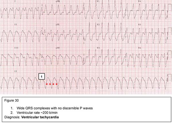
Ventricular tachycardia (VT)
VT is a rapid, wide QRS-complex tachycardia with a heart rate 150-250 beat/min. It is a serious condition that may result from drug toxicity (digoxin), myocarditis or severe metabolic derangement. It should be treated promptly with synchronized DC cardioversion if the patient is hemodynamically unstable. Stable VT may be treated with IV lidocaine infusion. Oral amiodarone may be used for outpatient management.
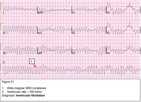
Ventricular Fibrillation (VF)
VF is a serious and terminal cardiac rhythm characterized by irregular, wide bizarre shaped QRS complexes. It needs urgent treatment with unsynchronized DC cardioversion.