 Imagine you have a brain tumor and your doctor says surgery is the only way to save your life. Now also imagine that the doctor has a 3-D image of your brain and using that image to “practice” the surgery before the actual procedure.
That may be what the future holds. At UTMB, doctors training to become neurosurgeons are using 3-D images mixed with a dose of virtual reality to identify diseased portions of the brain and to perform surgery.
The unique 3-D set up sits discreetly in a lab in UTMB’s John Sealy Hospital and is one of about 10 such simulators in the nation.
Bringing neurosurgical training into the 21st century has been the mission of Jaime Gasco, M.D., ever since he became director of neurosurgery resident education for UTMB in late 2011.
“What we have going on here,” Gasco says, “is what everyone should be doing.”
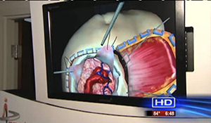
Once in the lab, Gasco leans forward, his attention fixed on the task in front of him. Above, on a large computer monitor, there’s a 3-D image of the top of a human head. Using essentially a very fancy joystick, Gasco “virtually” mimics the sensation of boring through bone and brain. There’s the whine of a drill as he approaches his goal.
“When I get to the ventricle, I should feel a pop as I enter this fluid-filled cavity,” he says. “There – I felt it. And it’s giving me a score of 13.7, which is a pretty good score.” Gasco leans back from the white arcade-game style cabinet where he has been working and removes his 3-D goggles.
“I learned to do surgery in the traditional way, which compared to this is sort of the caveman way of learning it,” Gasco says. “It looks like an easy thing to do, but you have to do a lot of these procedures before you get really good. And with this and our other simulators, our students can do that without ever touching a real patient.”
The lab also uses remarkably lifelike mannequins for surgery practice, including heads that contain texturally accurate brains and faithful representations of tumors and blood vessels that bleed simulated blood. “Mannequin simulators are good because you’re actually using the same instruments as in the operating room, so you build the same eye-hand coordination skills,” Gasco says.
He holds up a mannequin’s head. “We have mannequins where you can insert real endoscopes through the head and see tumors and fake blood vessels on camera – pretty amazing and remarkable things.”
Gasco and the residents recently took the simulations to a new level when they performed procedures on the mannequins in one of the operating rooms at John Sealy Hospital.
According to Gasco, his students appreciate the chance to work with such cutting-edge tools – although, never having had to learn the old-fashioned way, they probably don’t know how good they have it. It doesn’t bother him, though.
“In an academic institution, we have an obligation to make our residents better than we are,” he says. “That’s why I’m doing this.”