Secondary lesions
Secondary lesions are those lesions that are characteristically brought about by modification of the primary lesion either by the individual with the lesion or through the natural evolution of the lesion in the environment.
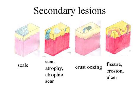
| Atrophy: localized shrinking of the skin which results in paper-thin, wrinkled skin with easily visible vessels. Results from loss of epidermis, dermis or both. Dermal atrophy manifests as a depression in the skin which can occur secondary to intralesional steroid injections. Epidermal atrophy manifests as thin almost transparent skin; may not retain normal skin lines which can occur secondary to topical steroid use. |
| Crust: occurs from dried exudate overlying and impaired epidermis. The exudate can be composed of blood, serum, or pus. e.g impetigo, epidermolysis bullosa. |
| Erosion: intraepithelial loss of epidermis, usually heals without scarring, moist, circumscribed, usually depressed lesion due to loss of all or part of the epidermis e.g. herpes simplex. |
| Fissure: linear, often painful breaks within the skin surface, as a result of excessive xerosis (dryness of skin). |
| Scale: occurs due to increased shedding or accumulation of stratum corneum as a result of abnormal keratinization and exfoliation (e.g. seborrheic dermatitis, postmaturity desquamation). Types of scale include pityriasiform which is branny and delicate, psoriaform which is thick, white and adherent, and icthyosiform which is fish-scale-like. |
| Scar: permanent fibrotic skin changes that develop as a consequence of tissue injury in which normal tissue is replaced by fibrous connective tissue at the site of injury to the dermis. Scars may be hypertrophic, atrophic, sclerotic or hard due to collagen proliferation. Reflects pattern of healing in the affected area. |
| Ulcer: full-thickness loss of the epidermis with damage into the dermis, heals with scarring (e.g. ulcerated hemangiomas, aplasia cutis congenita). |