


Transition to extra-uterine life
More Information on Fetal Circulation
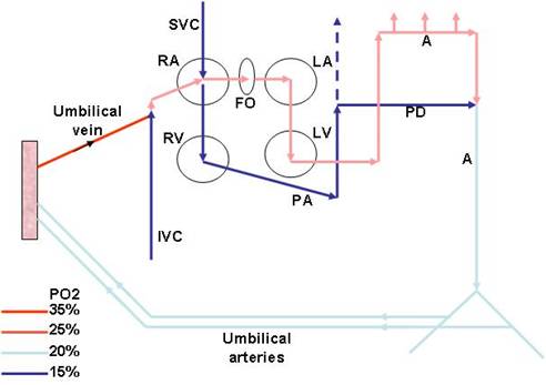
Diagram by A. Kalia
In the fetus, oxygenated blood from the placenta enters the umbilical vein and flows into the inferior vena cava (IVC) where it mixes with blood returning from the lower body.
From the IVC, blood enters the right atrium and preferentially flows across the open foramen ovale to the left atrium; this blood is then ejected by the left ventricle into the aorta and supplies the coronary arteries and the upper body, including the carotids.
Blood that is returned from the upper body by the superior vena cava is preferentially directed to the right ventricle and ejected into the pulmonary artery. The pulmonary vascular resistance in the fetus is high, and the most of this blood blood is shunted across the ductus arteriosus into the aorta and travels to the splanchnic area and the lower body. Much of this blood is routed via the umbilical arteries to the placenta for reoxygenation.
Onset of respiration:
Prior to birth, the lungs are filled with fluid and receive only 10 to 15% of the total cardiac output. With the onset of respiration, there is expansion of the lungs with air, a sharp decline in pulmonary vascular resistance, and an increase in pulmonary blood flow. Within the first minutes of life, much of the fluid in the lungs is removed by the lymphatics and by the thoracic squeeze resulting from vaginal delivery. Increase in the pO2, a decline in the pCO2, increased in pH and release of bradykinin and prostaglandins also contribute pulmonary arterial dilatation at birth. Pulmonary blood flow increases 8-10 fold, as does the volume of blood returning to the left heart, resulting in increased ventricular output.
Cardiovascular changes:
Before birth, distribution of left atrial blood between the tricuspid valve and the foramen ovale means that the two ventricles act in parallel. For the heart to function as two pumps in series - the pulmonary circulation and the systemic circulation - direct communications between the right and left sides of the circulation must be closed.
After the blood flow from the placenta is removed from the circulation, blood flow to the right atrium decreases. At the same time, blood flow to the left atrium increases because of increased venous return from the pulmonary circulation. The increase in left atrial pressure relative to right atrial pressure closes the valve-like flap overlying the foramen ovale in the left atrium.
The ductus arteriosus contains smooth muscle that is maintained in a relaxed state by the action of prostaglandin E1. After birth, serum levels of prostaglandin E1 fall and in most term infants, functional closure of the ductus occurs within the first 12 hours of life. If the ductus remains open, the flow through it will be reversed because of the increase in left ventricular output and aortic pressure. A patent ductus arteriosus leads to excessive pulmonary blood flow and may result in right heart failure. In this situation, indomethacin, an anti-prostaglanding agent, is used to try to achieve closure of the ductus.
On the other hand, certain congenital cardiac malformations, such as pulmonary atresia, require patency of the ductus arteriosus to maintain pulmonary blood flow. In this situation, prostaglandins may be infused intravenously to keep the ductus open until surgery can be performed.
In summary, prior to birth, the placenta is the conduit that provides oxygen and nutrition to the fetus. In the fetal circulation, pulmonary vascular resistance is high and right ventricular output is shunted across the ductus arteriosus into the systemic circulation, bypassing the lungs. At birth, dramatic changes occur in the fetal circulation that allow transition to a lung-based gas exchange.
Pulmonary vascular resistance falls and the ductus arteriosus closes. Right ventricular output now circulates through the lungs before entering the systemic circulation via the left atrium.
Changes also occur in other organ systems. Availability of oxygen from the lungs is better than from the placenta. Enzyme systems are activated in the liver. For example, glucoronyl transferase activation is essential for conjugating the bilirubin produced from the disintegrating RBCs. The kidneys assume responsibility for fluid and electrolyte homeostasis. The immune system has to mature.
Quickcheck: Transition to Extra-uterine life
![]()
Please select true or false

