Bo Chen, PhDAssistant Professor

Room 4.212D Research Building 17
Route: 0620 | Tel: (409) 747-2214 | bochen1@utmb.edu
Research Experts
Education and Training
B.Sc. Biological science, University of Birmingham, Birmingham, U.K.
M.Sc. Biomedical science, Durham University, Durham, U.K.
Ph.D. Applied biomedical science, University of Reading, Reading, U.K.
Postdoctoral training, Boston Children’s Hospital, Harvard Medical School, Boston, U.S.
Research Overview
Our lab studies how spinal cord injury changes surviving neurons and the circuits that connect the brain and spinal cord. We investigate why these circuits fail, how harmful cellular changes develop, and how targeted interventions can restore function. Our research focuses on movement, bladder control, and neuropathic pain. We combine quantitative three-dimensional imaging, circuit mapping and manipulation, and functional studies in experimental models to connect cellular mechanisms with recovery. inal cord.
Restoring spinal circuit function and protecting surviving neurons
During my postdoctoral training in Zhigang He’s laboratory, Harvard Medical School, we showed that enhancing KCC2, a neuronal chloride transporter, function in spinal inhibitory interneurons can reactivate dormant relay pathways and mprove stepping after spinal cord injury in mice. This work demonstrated that targeting dysfunctional circuits can enable spared connections to support recovery. Our lab now investigates how KCC2regulation changes across neuronal populations after injury and how these changes affect circuit function.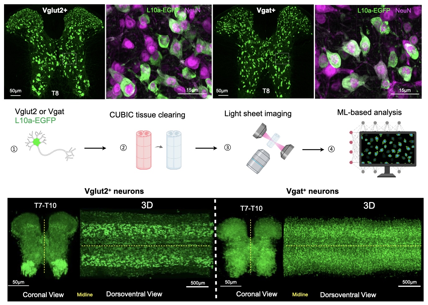
Restoring brain–spinal communication for bladder control
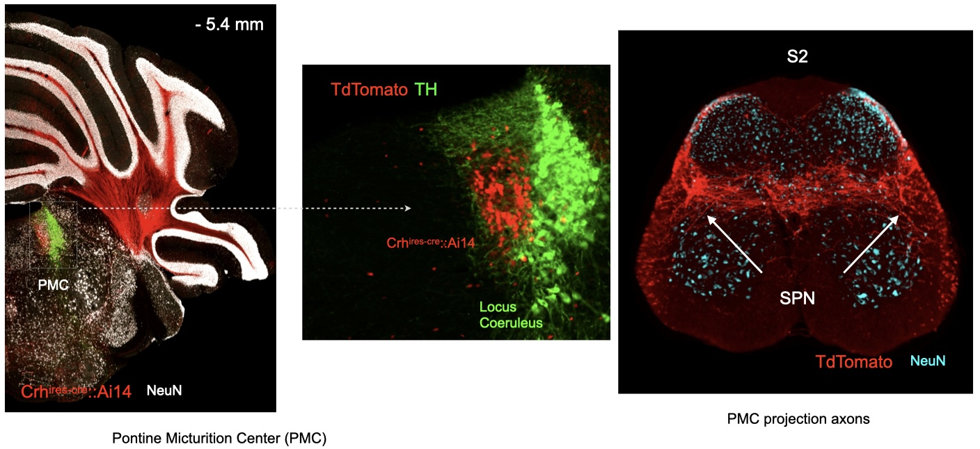 Loss of bladder control is a major consequence of spinal cord injury. Normal urination requires coordinated activity between the brain and spinal circuits that control bladder contraction and urethral sphincter relaxation. Spinal cord injury disrupts this communication, impairing both voluntary control and effective bladder emptying.We investigate how descending signals from the pontine micturition center (PMC) engage spinal circuits and how these connections change after injury. By mapping and testing these pathways, we aim to identify circuit mechanisms that can be targeted to restore coordinated bladder function and voluntary control.
Understanding and targeting circuits that drive neuropathic pain
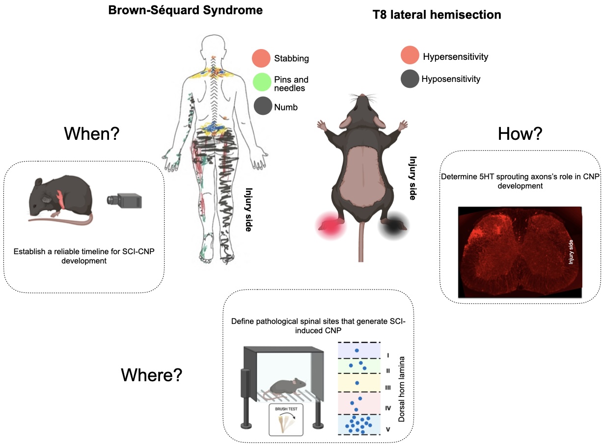 Using a spinal cord hemisection model that reproduces key sensory features of Brown-Séquard syndrome, we examine how descending pathways and spinal circuits contribute to abnormal pain processing. By identifying and testing the athways involved, we aim to develop strategies to prevent neuropathic pain and reduce pain after it has become established.
Selected Publications: