The Neurodevelopment Core’s mission is to empower investigators with physiologically relevant platforms that bridge basic discovery and clinical translation in neurodevelopmental and neurodegenerative disease research.
The Human Brain Organoids and Brain-on-a-Chip (BOC) Core at the Moody Brain Health Institute provides state-of-the-art, human stem cell–derived models that replicate key aspects of the human brain in a laboratory setting. By combining induced pluripotent stem cell (iPSC)–derived organoids with microphysiological systems (MPS), the Core equips MBHI and UTMB investigators with powerful, human-relevant tools to explore brain development, neurodegeneration, therapeutic discovery, and drug delivery.
These next-generation approaches reduce reliance on animal models and align with FDA and NIH initiatives promoting New Approach Methodologies (NAMs) to better assess safety and efficacy in preclinical research.
Overview
- Human Brain Organoids are 3D “mini-brains” generated from iPSCs reprogrammed from human blood or skin cells. They reproduce many features of the human brain—neuronal differentiation, synaptic connectivity, and glial interactions—allowing researchers to study disease mechanisms and therapeutic responses in a patient-specific context.
- Brain-on-a-Chip (BOC) systems recreate functional neural circuits using human neurons, glia, and microfluidic technology. These platforms enable real-time monitoring of neuronal network activity, neurovascular interactions, and cell-to-cell communication, advancing studies of brain injury, repair, and drug delivery.
Together, these innovative platforms form a transformative research resource for MBHI investigators—bridging discovery and translation to accelerate innovation, collaboration, and progress in brain health.
Key Services & Capabilities
Organoid & Brain-on-Chip (BOC) Development
Custom generation of cerebral and hippocampal organoids from human iPSCs, along with fabrication of advanced BOC systems for modeling defined neural circuits and disease-relevant pathways.
Advanced Cellular Modeling
Integration of specialized cell populations—including microglia—to study neuroimmune interactions, neurodevelopmental mechanisms, and early drivers of neurodegeneration.
Training & Technical Support
Hands-on training, consultation, and experimental design support for organoid culture, BOC assembly, assay development, and workflow optimization.
Functional & Structural Analysis
Access to MEA recordings, permeability assays, leukocyte infiltration models, synaptosome assays, and advanced imaging platforms such as high-resolution confocal and two-photon microscopy. Support includes fixation, sectioning, immunostaining, imaging, and 3D reconstruction.
Collaborative Research Opportunities
Cross-lab partnerships to develop new experimental models, accelerate discovery, and support translational neuroscience initiatives.
Current Research Areas
Microglia-Enriched Organoids & BOC Models
Developing systems that incorporate microglia to explore neuroimmune contributions to brain development and disease.
Tau Oligomer Studies in AD & NDAN
Evaluating how tau oligomers from Alzheimer’s disease (AD) and non-demented AD pathology (NDAN) brains affect neuronal and glial integrity.
Neuronal Network Mapping
Using MEA recordings to characterize functional neuronal activity in organoid systems.
Synaptic & Glial Pathology Assessment
Investigating synaptic structure and glial dysfunction through immunofluorescence, synaptosome isolation, high-resolution imaging, and live-cell microscopy.
Integrative Organoid–BOC Platforms
Combining organoid and BOC technologies to advance translational neurodegeneration research and therapeutic discovery. immunostaining, imaging, and 3D reconstruction.
Collaborative Research Opportunities
Cross-lab partnerships to develop new experimental models, accelerate discovery, and support translational neuroscience initiatives.

Maria-Adelaide Micci is a Professor of Anesthesiology at UTMB and leads neurodevelopmental research at the Moody Brain Health Institute. Her work focuses on neurogenesis, exosome signaling, and patient-derived brain organoids and chips to study mechanisms of brain injury and neurodevelopmental disorders.

Anna Fracassi is an Assistant Professor of Neurology and neuroscientist at UTMB. Her research focuses on how microglia and autophagy influence neuroinflammation, resilience, and cognition in Alzheimer’s disease, aiming to uncover therapeutic targets to slow neurodegeneration and promote brain health.

Elvis Cuevas is an Assistant Professor of Neurology and Research Fellow of the Moody Brain Health Institute at UTMB. She leads the Neurovascular Research Cuevas Laboratory, specializing in blood-brain barrier biology and brain-on-a-chip technologies to advance translational neuroscience.

Zahra Kolahchi earned her medical and public health degrees from Tehran University of Medical Sciences and is a Postdoctoral Research Fellow at UTMB. Her work focuses on the blood–brain barrier in Alzheimer’s disease, with research interests in dementia and cognitive neurology.

Kevin earned his PhD in Neuroscience from UTMB, where he investigated how hippocampal neurogenesis becomes disrupted in Alzheimer’s disease. He is now a Postdoctoral Fellow at UTMB, focusing on the development of advanced human brain organoid models to study the mechanisms underlying pathological tau propagation in Alzheimer’s disease and related tauopathies. His research interests include neurodegeneration, stem cell–based modeling, and the cellular pathways that drive disease progression.

Wen-Ru Zhang is a Research Associate II in the Neurodevelopmental Systems Lab at the Moody Brain Health Institute, bringing over three decades of experience in neuroscience and molecular biology. She specializes in protein and gene expression analyses and supports advanced brain model development to advance translational neurobiology and regenerative medicine

Yaping Zheng is a Research Associate II in the Neurodevelopment Core at the Moody Brain Health Institute, where she specializes in generating and maintaining human cerebral organoids from induced pluripotent stem cells. With over two decades of experience at UTMB, she advances 3D brain model development to support translational research in neurodevelopment disorders.
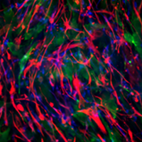
Exploring Brain Organoids
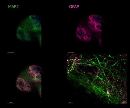
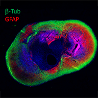
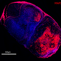
After more than 80 days in culture, this human brain organoid demonstrates advanced development of key brain cell types — neurons and astrocytes — that are essential for modeling brain function and disease. The images show expression of:
- MAP2 (green): a neuronal marker highlighting the extensive network of developing neurons.
- GFAP (pink): an astrocytic marker showing supportive glial cells distributed across the organoid surface.
The merged images reveal intricate interactions between neurons and astrocytes, closely mimicking the architecture of the human brain. The accompanying 3D video rendering provides a detailed visualization of the organoid’s structure, allowing viewers to explore its cellular complexity and spatial organization. Together, these tools illustrate how MBHI’s Neurodevelopment Core is advancing the use of 3D human brain models to study neurodevelopment, neurodegeneration, and brain repair.
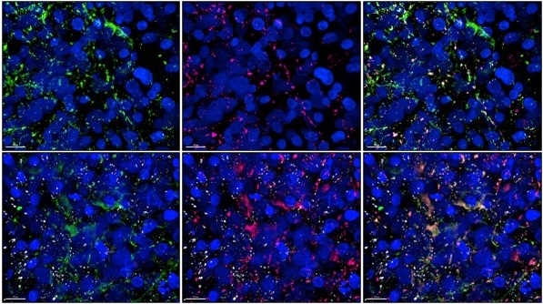
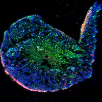
After more than 80 days in culture, 3D human brain organoids made from stem cells were exposed to tau proteins linked to Alzheimer’s disease. Within 24 hours, these tau proteins (shown in white) began forming clusters inside the organoids, while untreated samples showed no buildup.
The images show:
- Green: normal tau proteins forming networks inside brain cells.
- Pink: abnormal, disease-related tau proteins beginning to clump together.
- Merged image: overlap of both types of tau, showing how exposure triggers aggregation similar to what occurs in neurodegenerative diseases.
These findings demonstrate that MBHI’s organoid models can replicate key early processes of Alzheimer’s pathology, providing a powerful system for studying how toxic proteins spread and interact in the human brain.
Understanding the Brain-on-a-Chip Model
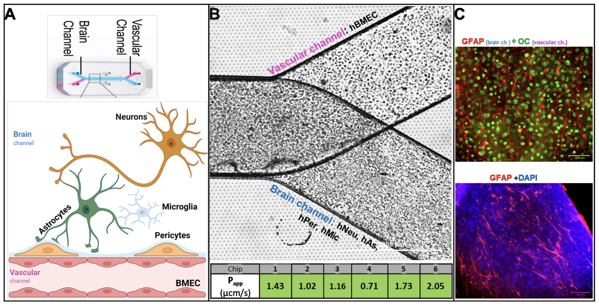
Our Brain-on-a-Chip (BOC) system recreates two key parts of the human brain environment using tiny, parallel channels separated by a thin, breathable membrane.
- The “vascular channel” is lined with human brain blood vessel cells. This models the blood-brain barrier (BBB)—the protective layer that controls what enters and leaves the brain.
- The “brain channel” contains a mix of brain cells, including neurons, astrocytes, microglia, and pericytes. Together, these cells represent the brain tissue side of the barrier.
Because the two channels are kept separate, researchers can adjust conditions in each one independently. This makes it possible to study how brain cells and blood-vessel cells communicate, how each side responds to disease-like conditions, and how treatments may affect the brain.
We also control the flow of fluids through the channels to mimic real-life brain and blood-vessel environments.
Panel B: When the blood-vessel cells form a strong, tight layer, we see low permeability values (Papp < 2). This tells us the model has successfully formed a working blood-brain barrier.
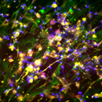
Panel C: Fluorescent labeling helps visualize the cells in each channel—blood-vessel cells glowing in the vascular channel and astrocytes glowing in the brain channel—confirming that each cell type is in the correct location.


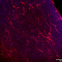
Representative immunofluorescence image of the Blood-Brain Barrier (BBB) cultured on the Emulate Brain-Chip.
Primary human brain microvascular endothelial cells in the bottom (vascular) channel are stained for occludin (green), showing tight junction formation.
Primary human astrocytes in the top (parenchymal) channel are stained for GFAP (red). Nuclei in both channels are counterstained with DAPI (blue).