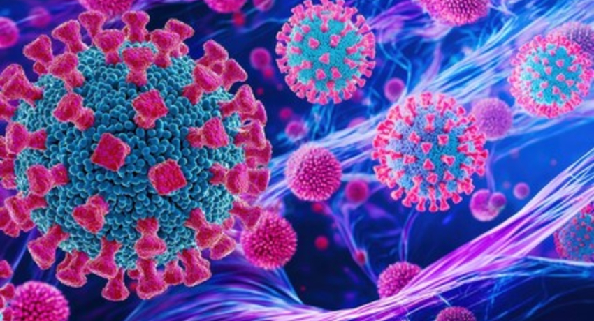
Researchers at The University of Texas Medical Branch (UTMB) have identified two SARS-CoV-2 proteins that play a major role in helping the virus suppress the body’s early immune defenses, offering new insight into how COVID-19 causes infection and disease.
In a new study, scientists examined how SARS-CoV-2 uses a collection of viral proteins to interfere with the innate immune response—the body’s first line of defense against infection. While previous studies suggested that several viral proteins could dampen immune activity, most examined proteins individually in different experimental systems, making it impossible to directly compare their relative importance during actual viral infection.
To answer that question, the research team engineered 12 recombinant versions of SARS-CoV-2, each carrying mutations in a different viral protein. The researchers then compared how the immune system responded to each modified virus. The study revealed that among 12 tested proteins, mutations in two—NSP1 and NSP15—triggered significantly stronger antiviral immune responses than the original virus, with nine of the 12 proteins showing some immune-suppressing activity.
Further analysis showed that weakening NSP1 or NSP15 led to increased activation of genes involved in antiviral defense, immune signaling, and immune cell communication. The altered viruses also promoted greater maturation of dendritic cells, specialized immune cells that help coordinate the body’s adaptive immune response.
When tested in an animal model, viruses with mutations in NSP1 or NSP15 replicated less efficiently in the respiratory tract, suggesting these proteins contribute to the virus’ ability to establish infection and evade immune detection.
Using single-cell RNA sequencing, the researchers also identified broad changes in immune cell activation patterns in the lungs of infected mice, including effects on myeloid and lymphoid immune cell populations.
 "We built 12 distinct infectious viruses and tested them side by side under identical conditions, in multiple experimental systems," said senior author Alexander Bukreyev, PhD, head of the Laboratory of Viral Pathogenesis and Vaccine Development at UTMB. “That level of scale has not been done before for this virus, and the map it produces of how SARS-CoV-2 hijacks early immune defenses is exactly what is needed to design effective interventions."
The findings demonstrate that SARS-CoV-2 relies on multiple, overlapping mechanisms to suppress immune activity and avoid detection. The study highlights NSP1 and NSP15 as especially important contributors to immune evasion and viral pathogenesis.
 “Our findings show that SARS-CoV-2 uses multiple proteins to interfere with the body’s early immune defenses, but NSP1 and NSP15 appear to play especially important roles,” said lead researcher Fuchun Zhou, PhD, a research scientist in the department of pathology at UTMB. “By weakening those proteins, we saw a much stronger antiviral response and reduced viral replication, which helps us better understand how the virus establishes infection and how future therapies might disrupt those immune-evasion strategies.
The research provides a ranked understanding of which SARS-CoV-2 proteins are most critical to manipulating the immune system, helping prioritize which proteins should be targeted in the development of future antiviral therapies and vaccine strategies.
Additional authors on this research: Sivakumar Periasamy, Ruben Soto Acosta, Aarti Tripathi, Kritika Kedarinath, Philipp A. Ilinykh, and Shailendra Chauhan, Department of Pathology and the Galveston National Laboratory at the University of Texas Medical Branch; Haiping Hao and Steven G. Widen, Department of Biochemistry and Molecular Biology, University of Texas Medical Branch; Nathaniel D. Jackson, Chengjin Ye, and Luis Martinez-Sobrido, Texas Biomedical Research Institute, San Antonio; and Wan Sze Cheng, German Nudelman, Elena Zaslavsky, and Stuart C. Sealfon, Department of Neurology, Icahn School of Medicine at Mount Sinai, New York