Comprehensive pancreatic and biliary care
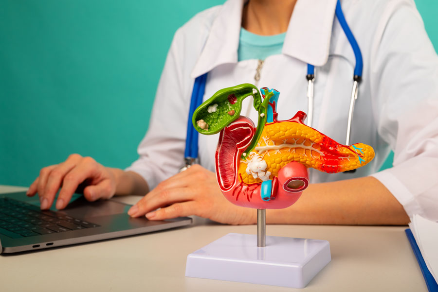
Pancreatic and biliary disorders can cause significant digestive discomfort and serious health complications if left untreated. At UTMB Health, our specialists use advanced diagnostic and therapeutic techniques to evaluate and treat complex pancreatic and bile duct conditions. You can expect nothing but precise, minimally invasive care focused on restoring your digestive function and improving your long-term health.
Conditions We Treat
Bile Duct Cancer (Cholangiocarcinoma)
Overview:
Bile duct cancer, or cholangiocarcinoma, is a rare but aggressive cancer that develops in the bile ducts connecting the liver and gallbladder to the small intestine. It typically affects older adults and may cause jaundice or abdominal discomfort. Early detection is challenging, but advanced imaging and endoscopic techniques help guide treatment options, which may include surgery, chemotherapy, and targeted therapies.
Common Symptoms:
- Jaundice
- Abdominal pain
- Itchy skin
- Dark urine, pale stools
- Weight loss and fatigue
Treatments & Procedures:
- Surgical removal of tumors (when possible)
- Biliary stenting to relieve obstruction
- Chemotherapy and radiation therapy
- Targeted or immunotherapy (for advanced cases)
- Palliative care for symptom control
Bile Duct Stones (Choledocholithiasis)
Overview:
Bile duct stones occur when gallstones migrate from the gallbladder into the common bile duct, blocking bile flow. This can lead to jaundice, infection, or pancreatitis. The condition most often affects adults with a history of gallstones. Prompt diagnosis and removal of the stones are essential to restore bile drainage and prevent serious complications.
Common Symptoms:
- Upper abdominal pain
- Jaundice (yellowing of skin and eyes)
- Dark urine and pale stools
- Fever and chills
- Nausea or vomiting
Treatments & Procedures:
- Endoscopic retrograde cholangiopancreatography (ERCP) for stone removal
- Antibiotics for infection
- Laparoscopic or open surgery (if ERCP not possible)
- Follow-up imaging to prevent recurrence
Bile Duct Strictures or Obstructions
Overview:
Bile duct strictures or obstructions occur when the ducts carrying bile become narrowed or blocked, often from injury, gallstones, inflammation, or tumors. This disrupts bile flow and may cause jaundice, infection, or liver damage. The condition requires imaging and endoscopic evaluation to identify the cause and restore normal drainage through minimally invasive or surgical procedures.
Common Symptoms:
- Jaundice
- Dark urine and light stools
- Itchy skin
- Abdominal pain
- Fever (if infection present)
Treatments & Procedures:
- ERCP with stent placement or dilation
- Percutaneous biliary drainage
- Surgical reconstruction (for severe or recurrent cases)
- Antibiotics for infection
- Monitoring of liver function
Biliary Colic
Overview:
Biliary colic is a type of pain caused by temporary blockage of the bile duct, usually from a gallstone. The obstruction prevents bile from flowing normally, triggering severe, intermittent pain. It often follows a fatty meal and may last from minutes to hours. While episodes may resolve on their own, recurring pain often indicates gallbladder disease requiring further evaluation or surgery.
Common Symptoms:
- Sudden, steady pain in upper right abdomen
- Pain radiating to back or shoulder
- Nausea and vomiting
- Pain after fatty meals
Treatments & Procedures:
- Pain management and dietary changes
- Evaluation for gallstones via ultrasound
- Laparoscopic cholecystectomy to prevent recurrence
Exocrine Pancreatic Insufficiency
Overview:
Exocrine pancreatic insufficiency (EPI) occurs when the pancreas cannot produce enough digestive enzymes to properly break down food. It is often caused by chronic pancreatitis, cystic fibrosis, or pancreatic surgery. EPI leads to malnutrition and gastrointestinal symptoms. With appropriate enzyme replacement and dietary management, patients can restore normal digestion and improve nutrient absorption and quality of life.
Common Symptoms:
- Chronic diarrhea or loose stools
- Greasy, foul-smelling stools
- Weight loss
- Bloating and gas
- Fatigue or weakness
Treatments & Procedures:
- Pancreatic enzyme replacement therapy (PERT)
- Nutritional supplementation (fat-soluble vitamins)
- High-calorie, low-fat diet adjustments
- Treatment of underlying pancreatic disease
- Regular monitoring of nutritional status
Gallbladder Inflammation (Cholecystitis)
Overview:
Cholecystitis is inflammation of the gallbladder, usually caused by gallstones blocking bile flow. It can lead to infection and severe pain requiring urgent medical attention. The condition commonly affects adults with gallstones and may recur without treatment. Early diagnosis and management are critical to prevent complications such as gallbladder rupture or widespread infection.
Common Symptoms:
- Severe right upper abdominal pain
- Fever and chills
- Nausea or vomiting
- Tenderness over the gallbladder area
- Pain after meals
Treatments & Procedures:
- Hospitalization for intravenous fluids and antibiotics
- Pain management
- Laparoscopic cholecystectomy (definitive treatment)
- Drainage procedure for high-risk patients
Gallstones
Overview:
Gallstones are hardened deposits that form in the gallbladder from imbalances in bile components. They may be small and asymptomatic or cause pain if they block bile flow. Gallstones are more common in women, older adults, and those with obesity or rapid weight loss. Treatment depends on symptom severity and may include dietary changes, medications, or surgical removal of the gallbladder.
Common Symptoms:
- Sudden right upper abdominal pain
- Pain after eating fatty foods
- Nausea or vomiting
- Back or shoulder pain
- Bloating or indigestion
Treatments & Procedures:
- Observation for asymptomatic cases
- Dietary modification (low-fat diet)
- Medications to dissolve small stones (rare)
- Laparoscopic cholecystectomy (gallbladder removal)
Pancreatic Cancer
Overview:
Pancreatic cancer forms in the pancreas, the organ that helps with digestion and blood sugar control. It’s often diagnosed late because early symptoms are subtle. Risk factors include smoking, diabetes, and family history. Doctors aim to remove tumors safely while supporting digestion, blood sugar, and overall health. Recovery and follow-up care are key to improving outcomes.
Common Symptoms:
- Upper abdominal pain
- Unexplained weight loss
- Jaundice (yellowing of skin/eyes)
- Nausea and loss of appetite
- Back pain
Treatments & Procedures:
- Whipple procedure (pancreaticoduodenectomy)
- Distal or total pancreatectomy
- Chemotherapy
- Radiation therapy
- Supportive care for digestion and blood sugar
Pancreatic Cysts or Pseudocysts
Overview:
Pancreatic cysts and pseudocysts are fluid-filled sacs that form on or within the pancreas. Pseudocysts often develop after pancreatitis, while true cysts may be benign or precancerous. Most are discovered incidentally during imaging. Management depends on size, symptoms, and cancer risk, ranging from observation to drainage or surgical removal for large or suspicious cysts.
Common Symptoms:
- Upper abdominal fullness or pain
- Nausea or vomiting
- Bloating
- Early satiety
- Fever (if infection occurs)
Treatments & Procedures:
- Imaging surveillance for small, asymptomatic cysts
- Endoscopic or percutaneous drainage
- EUS-guided fine-needle aspiration (for diagnosis)
- Surgical removal of complex or precancerous cysts
Pancreatitis (Acute and Chronic)
Overview:
Pancreatitis is inflammation of the pancreas, occurring as either an acute episode or a chronic, progressive condition. Common causes include gallstones, alcohol use, or high triglycerides. Acute cases may resolve with supportive care, while chronic pancreatitis can lead to permanent damage and enzyme deficiency. Early diagnosis and ongoing management help prevent complications and preserve digestive function.
Common Symptoms:
- Severe upper abdominal pain radiating to the back
- Nausea and vomiting
- Fever
- Bloating or tenderness
- Fatty, foul-smelling stools (chronic)
Treatments & Procedures:
- Fasting and intravenous fluids for acute cases
- Pain management
- Endoscopic or surgical removal of gallstones
- Pancreatic enzyme replacement (chronic cases)
- Lifestyle changes and alcohol cessation
Services & Procedures
Bile Duct Leak Stenting (Post-Surgery)
Overview:
Bile duct leak stenting is an endoscopic treatment used to seal bile leaks that can occur after gallbladder or liver surgery. A small stent is placed through endoscopic retrograde cholangiopancreatography (ERCP) to divert bile flow and allow the leak site to heal naturally, avoiding open surgery.
Conditions Treated:
- Bile leaks after cholecystectomy or liver resection
- Post-traumatic bile duct injuries
- Minor bile duct disruptions or strictures
Before the Procedure:
You’ll be asked to fast for 6-8 hours before the procedure, which is performed under sedation or anesthesia. Blood tests and imaging (CT, MRCP) may be performed to locate the leak.
After the Procedure:
most patients recover quickly and can resume a normal diet after observation. Mild bloating or nausea may occur. The stent is usually removed or exchanged after several weeks once healing is confirmed with imaging or repeat ERCP.
Capsule Endoscopy
Overview:
Capsule endoscopy is a noninvasive diagnostic procedure that uses a small, pill-sized camera to capture images of the digestive tract, especially the small intestine, which is difficult to view with standard endoscopy. The capsule transmits thousands of pictures to a recording device as it passes naturally through the GI tract, helping identify bleeding, inflammation, or other abnormalities.
Conditions Evaluated:
- Unexplained gastrointestinal bleeding
- Crohn’s disease of the small bowel
- Celiac disease (to assess mucosal damage)
- Small bowel tumors or polyps
- Unexplained abdominal pain or anemia
Before the Procedure:
Patients typically fast overnight and may be instructed to use a bowel prep for clear imaging. Metal objects or electronic devices near the recorder should be avoided. The capsule is swallowed with water, and normal activity can usually continue during the recording period.
After the Procedure:
The capsule passes naturally in the stool, usually within 24-48 hours. Patients return the recording device for image review. Normal diet and medications can resume once instructed. Rarely, the capsule may become lodged if there is a bowel stricture, so patients should report any persistent pain or failure to pass the capsule.
Endoscopic Drainage of Fluid Collections or Cysts
Overview:
Endoscopic drainage uses ultrasound-guided tools to access and drain abnormal fluid collections, such as pancreatic pseudocysts or abscesses, through the stomach or intestinal wall. A stent or catheter may be placed for continued drainage. This minimally invasive approach helps avoid open surgery and speeds recovery.
Conditions Treated:
- Pancreatic pseudocysts
- Walled-off pancreatic necrosis
- Abdominal abscesses near GI tract
- Post-surgical fluid collections
Before and After the Procedure:
You’ll be expected to fast for several hours before the procedure. Imaging (CT or MRI) confirms the collection’s size and location. The procedure is done under deep sedation or anesthesia. After the procedure, some abdominal soreness or bloating may occur. Antibiotics are often prescribed. Follow-up imaging ensures the fluid has resolved. The stent is removed later once drainage is complete.
Endoscopic Mucosal Resection (EMR) and Endoscopic Submucosal Dissection (ESD)
Overview:
EMR and ESD are advanced endoscopic procedures used to remove abnormal or early-stage cancerous tissue from the lining of the digestive tract without surgery. EMR removes smaller, superficial lesions in pieces, while ESD allows en bloc (single-piece) removal of larger or deeper lesions. Both help preserve healthy tissue and reduce the need for open surgery.
Conditions Treated:
- Early gastric, esophageal, or colorectal cancer
- Large or precancerous polyps
- Barrett’s esophagus with dysplasia
- Superficial mucosal tumors
Before the Procedure:
You'll be expected to fast for several hours and may need bowel prep for lower GI procedures. Anticoagulants or blood thinners are paused under medical supervision. The procedure is done under sedation or anesthesia.
After the Procedure:
Mild discomfort, bloating, or sore throat can occur. Diet advances gradually from liquids to solids as tolerated. Avoid NSAIDs or anticoagulants until cleared. Rare complications include bleeding or perforation, which are usually managed endoscopically.
Endoscopic Retrograde Cholangiopancreatography
Overview:
Endoscopic retrograde cholangiopancreatography (ERCP) is a specialized endoscopic procedure used to diagnose and treat problems in the bile ducts, gallbladder, and pancreas. It combines endoscopy (using a flexible tube with a camera) with X-ray imaging.
During ERCP, a scope is guided through the mouth into the small intestine. A thin tube is then inserted into the bile or pancreatic ducts, and a contrast dye is injected to make these ducts visible on X-ray. This allows the doctor not only to see blockages or narrowing but also to treat them in the same procedure.
Conditions Treated:
- Bile duct stones (choledocholithiasis)
- Biliary strictures or obstructions
- Blockages from tumors (pancreatic, bile duct, or gallbladder cancer)
- Leaks in the bile or pancreatic ducts (after surgery or injury)
- Some cases of cholangitis (infection of the bile ducts)
Before the Procedure:
- Fasting: You will need to stop eating and drinking for about 6–8 hours before the procedure.
- Medication review: Tell your doctor about blood thinners, diabetes medications, or allergies. Some medicines may need to be paused.
- Consent: The procedure, risks, and benefits will be explained; you will sign a consent form.
- IV placement: An IV line is placed for sedation or anesthesia.
- Sedation: Most patients receive conscious sedation or general anesthesia to stay comfortable and relaxed.
After the Procedure:
- Recovery: You’ll rest in a recovery area until the sedation wears off (usually 30–60 minutes).
- Escort needed: You will need someone to drive you home.
- Diet: Most patients can resume normal eating and drinking later the same day, unless instructed otherwise.
- Mild effects: Sore throat, bloating, or mild abdominal discomfort may occur.
- Follow-up: Your doctor will review results and any biopsy findings.
Endoscopic Ultrasound
Overview:
Endoscopic ultrasound (EUS) combines endoscopy (a thin flexible tube with a camera) and ultrasound imaging to create detailed pictures of the digestive tract and nearby organs. The scope is passed through the mouth or rectum, and an ultrasound probe at the tip provides high-resolution images from inside the body.
EUS can be used to both diagnose and treat various gastrointestinal conditions. As a diagnostic tool, it provides detailed imaging and enables tissue sampling. As a therapeutic tool, it offers minimally invasive alternatives to surgery for drainage, pain management, and targeted interventions. It’s commonly used to evaluate mediastinal lymphadenopathy of unknown origin, pancreatic masses, and biliary tract lesions.
Conditions Treated:
- Pancreatic cysts
- Pancreatitis (chronic and acute)
- Biliary strictures or obstructions
- Bile duct stones (choledocholithiasis)
- Primary sclerosing cholangitis
- Liver metastases
- Portal hypertension
- Gastrointestinal (GI) bleeding
- Unexplained abdominal pain or bloating
- Unexplained weight loss
- Colorectal cancer
Before the Procedure:
- Fasting: You’ll need to stop eating and drinking for about 6–8 hours before the test so your stomach is empty.
- Medication review: Your doctor will ask about blood thinners, diabetes medications, or allergies to anesthesia. Some medicines may need to be paused.
- Consent & prep: The care team will explain the procedure, risks, and benefits. You’ll sign a consent form.
- IV placement: An IV line will be placed for sedation or anesthesia.
- Sedation: Most patients receive conscious sedation or general anesthesia so you’re comfortable and drowsy.
After the Procedure:
- Recovery: You’ll rest in a recovery area until the sedation wears off (usually 30–60 minutes).
- Escort needed: Because of sedation, you’ll need someone to drive you home.
- Diet: Most people can resume normal eating and drinking later the same day, unless instructed otherwise.
- Mild effects: You may have a sore throat, bloating, or mild cramping for a day or so.
- Restrictions: Avoid driving, operating machinery, or making important decisions for 24 hours due to sedation.
- Follow-up: Your doctor will review results with you or schedule a follow-up appointment. Biopsy results may take several days.
Feeding Tube Placement
Overview:
Feeding tube placement (gastrostomy or PEG tube) is a procedure that delivers nutrition directly into the stomach when oral intake is inadequate or unsafe. In pancreatic and biliary disorders, it supports patients with severe malnutrition, pain, obstruction, or impaired digestion from conditions such as pancreatitis, pancreatic cancer, or biliary obstruction. It does not treat the underlying disease but helps maintain nutrition and strength during illness or recovery.
Conditions Treated/Evaluated:
- Chronic pancreatitis
- Severe acute pancreatitis (prolonged course)
- Pancreatic exocrine insufficiency
- Pancreatic cancer
- Pancreatic pseudocyst with poor oral intake
- Post-pancreatic surgery nutritional failure (e.g., Whipple recovery)
- Cholangiocarcinoma (bile duct cancer)
- Obstructive jaundice with cachexia/malnutrition
- Primary sclerosing cholangitis (advanced disease)
- Severe biliary obstruction with weight loss
- Post-biliary surgery recovery (e.g., hepaticojejunostomy)
Before the Procedure:
- You will not eat or drink for about 6–8 hours before the procedure.
- Blood tests may be done to check clotting and overall health.
- Your care team reviews medications and may pause blood thinners.
- An IV line is placed for fluids and sedation.
- The procedure and risks are explained, and consent is obtained.
After the Procedure:
- You are monitored for pain, bleeding, or complications.
- The feeding tube site is kept clean and dry.
- Feeding usually starts slowly within 12–24 hours if stable.
- Tube feeds are increased as tolerated.
- You are taught how to care for the tube and use it at home.
Neurolytic Celiac Plexus Block
Overview:
The neurolytic celiac plexus block (NCPB), often referred to as celiac plexus neurolysis (CPN) or a celiac axis block, is a specialized procedure used in pain management. It involves intentionally damaging specific nerves in the abdomen. This is done to alleviate severe pain, particularly pain caused by pancreatic cancer or other cancers affecting the upper gastrointestinal tract.
Conditions Treated:
- Severe pain from pancreatic cancer
- Chronic pancreatitis pain unresponsive to medication
- Other upper abdominal malignancies causing nerve-related pain
Before the Procedure
Patients fast for several hours. The procedure is performed under sedation or anesthesia, typically using endoscopic ultrasound or fluoroscopic guidance. Blood thinners may be paused beforehand.
After the Procedure:
Mild abdominal soreness, diarrhea, or transient blood pressure changes can occur but usually resolve quickly. Most patients experience significant pain relief within days. Activity can resume as tolerated, and follow-up visits assess symptom improvement and the need for additional treatments.
Placement of Stents in the GI Tract
Overview:
GI stent placement involves inserting a small mesh tube (stent) into a narrowed or blocked section of the esophagus, stomach, bile duct, or colon to keep it open. Performed endoscopically, it restores the passage of food or fluids and relieves obstruction caused by cancer, strictures, or scarring.
Conditions Treated:
- Esophageal or colorectal strictures
- Malignant obstructions (esophageal, biliary, duodenal)
- Post-surgical or inflammatory narrowing
- Biliary or pancreatic duct obstruction
Before and After the Procedure:
You’ll be expected to fast for 6-8 hours before the procedure. Sedation or anesthesia is used. Imaging or prior endoscopy helps plan stent size and placement. After the procedure, you’ll be monitored for pain, bleeding, or migration of the stent. Liquids are introduced first, followed by soft foods. Most patients can return to normal activity within a day.
POEM Procedure
Overview:
Peroral endoscopic myotomy, or POEM, is a minimally invasive endoscopic procedure to treat swallowing disorders caused by tight or spastic esophageal muscles. Using an endoscope inserted through the mouth, a small tunnel is created in the esophageal wall, and the inner muscle layer is cut (myotomy) to relax the esophagus and restore normal swallowing.
Conditions Treated:
- Achalasia
- Esophageal motility disorders (spasm, jackhammer esophagus)
- Zenker’s diverticulum (select cases)
Before and After the Procedure:
You'll be expected to fast overnight before the procedure and may receive antibiotics. Sedation or general anesthesia is used. Certain medications, such as anticoagulants, are paused per physician guidance. After the procedure, a clear liquid diet starts once swallowing is checked, then advances slowly. Temporary chest discomfort, sore throat, or bloating can occur. Proton pump inhibitors may be prescribed to prevent reflux.
Your Care Team
Each provider may treat different conditions. Click on a profile to see their areas of specialty.
General GI Providers
In some cases, our general gastroenterology specialists collaborate with UTMB Health's gastrointestinal surgeons as part of our advanced endoscopy services.
Frequently Asked Questions.
-
What are pancreatic and biliary disorders?
Pancreatic and biliary disorders include conditions that affect the pancreas, gallbladder, and bile ducts, organs that help digest food and regulate metabolism. Common conditions include gallstones, pancreatitis, bile duct obstructions, and pancreatic or biliary cancers.
-
What causes pancreatic and biliary disorders?
Causes range from gallstones and alcohol use to infections, inflammation, genetic factors, or tumors. Some disorders, like pancreatitis or bile duct obstruction, may result from gallstones blocking the flow of bile or pancreatic enzymes.
-
How are pancreatic and biliary conditions diagnosed?
UTMB Health specialists use advanced imaging and endoscopic techniques such as endoscopic ultrasound (EUS) and endoscopic retrograde cholangiopancreatography (ERCP) to accurately diagnose and treat pancreatic and biliary diseases. Blood tests, CT scans, and MRI may also be used.
-
What is ERCP, and when is it used?
ERCP stands for endoscopic retrograde cholangiopancreatography. It’s a minimally invasive procedure that combines endoscopy and X-rays to diagnose and treat problems in the bile and pancreatic ducts, such as gallstones, strictures, or blockages.
-
What’s the difference between acute and chronic pancreatitis?
Acute pancreatitis occurs suddenly and usually resolves with treatment, while chronic pancreatitis is long-term inflammation that can cause permanent damage and affect digestion. Both require medical management to prevent complications.
-
Can lifestyle changes prevent pancreatic and biliary disorders?
Yes. Maintaining a healthy diet, limiting alcohol intake, avoiding smoking, managing weight, and controlling cholesterol can lower the risk of gallstones, pancreatitis, and other related conditions.

All Gastroenterology Specialties
Our Locations
- 1005 Harborside Drive
6th Floor - M–F: 8 a.m.–5 p.m.
- (409) 772-0122
- (409) 747-0777
- Website
- 2240 Gulf Freeway South
Suite 2.110 - M–F: 8 a.m.–5 p.m.
- (832) 505-1600
- (281) 309-0147
- Website
- 2660 Gulf Freeway South
Entrance B - M–F: 8 a.m.–5 p.m.
- (832) 505-2350
- (281) 337-0719
- Website
- 17448 Highway 3
2nd Floor - M–F: 8 a.m.–5 p.m.
- (832) 505-1600
- (832) 905-6948
- Website
- 1715 South Friendswood Drive, 4th Floor
- M–F: 8 a.m.–5 p.m.
- (832) 505-1600
- (281) 482-3963
- Website
- 250 Blossom Street
2nd floor, Suite 230 - M–F: 8 a.m.–5 p.m.
- (832) 505-1600
- (832) 553-0292
- Website