When a scan reveals a suspicious spot on a patient’s lung, time matters. At The University of Texas Medical Branch (UTMB Health), artificial intelligence is helping clinicians identify those findings sooner and connect patients with the follow-up care they may need.
AI is now helping streamline the work of the Comprehensive Lung Cancer Screening Program at UTMB Health, including identifying lung nodules discovered unexpectedly on imaging performed for other medical reasons.
UTMB Health established the program in 2016 to screen patients who are at high risk for lung cancer and identify patients with incidentally found lung nodules who may require additional evaluation.
But according to Shawn Nishi, MD, medical director of the Comprehensive Lung Cancer Screening & Incidental Lung Nodule Program, the original process was time-consuming, sometimes taking weeks or even months. The screening team had to review records manually before identifying patients who needed follow-up, notifying them and their ordering providers, and scheduling additional testing or physician visits.
“By the time we navigated the care pathway for these patients, their time-critical follow-up window had often closed,” Nishi said. “We needed to replace our labor-intensive workflow with a faster, more sustainable process that prioritizes patient outcomes.”
AI automation improves follow-up process
Today, an artificial intelligence application is helping ensure patients receive more timely follow-up care.
“By implementing AI, we've transformed a highly manual, time-consuming workflow. We can now identify patients following screening exams and accelerate their path to a definitive outcome in a fraction of the time,” Nishi said.
AI is also helping identify incidentally discovered lung nodules. Any nodule, whether found on a lung cancer screening exam or another imaging study involving any portion of the lungs, is now flagged for review by the patient navigator.
Much of the chart review process, previously the most time-consuming part of the workflow, has been automated, allowing patient navigators to focus on helping patients understand and complete their next steps.
Nishi said the program has used several AI tools over the last few years.
“The current system, developed in collaboration with Bunkerhill, reviews radiology reports for patients undergoing lung cancer screening exams,” Nishi said.
Human oversight is key
While AI works in the background, people remain at the center of the process.
“Amanda Rodriguez is our patient navigator who is passionate about her work. She goes through these scans and makes sure that the AI model is working in the background and everything we intend in our process is followed,” Nishi said.
Rodriguez can view a dashboard utilizing data from patients’ electronic medical records to calculate a validated lung cancer risk score for patients with scan abnormalities. This enables her to efficiently prioritize follow-up care.
“She can go through every day and say, ‘There are five people today who haven’t followed up, and they have a really high risk for developing lung cancer based on this scan. I really want to focus on them first,’” Nishi said.
Nishi emphasized that AI is an assistant, not a decision maker.
The benefits of incidental screening
Because the protocol includes imaging involving any portion of the chest performed at UTMB Health, the program can flag pulmonary nodules discovered during imaging ordered for other reasons. Roughly two-thirds of the nodules identified through the program have been found in patients who were not undergoing lung cancer screening.
For Nishi, one patient illustrates the potential impact.
“I had a patient, in her 20s, who had a motor vehicle accident,” Nishi said. “She got a scan, and we found a nodule incidentally. We performed a biopsy and she did have lung cancer and ultimately underwent resection for cure."
When the patient was questioned about her family history, she named seven or eight relatives who had been diagnosed with cancer.
“She ended up having a hereditary disease, which made it important for her family members to be tested for the same disease,” Nishi said. "That’s a prime example of somebody we would never have caught if not for her motor vehicle accident.”
The case demonstrates how findings unrelated to the original reason for an imaging exam can lead to further evaluation and, in some cases, potentially life-changing care.
AI with a human touch
For patients who have an abnormal finding, technology is only one part of the process. The follow-up clinical visit provides important context and an opportunity for patients to talk directly with their care team.
“Fear often stems from the unknown,” Nishi said. “That’s why a clinic visit is so powerful. It gives people the opportunity to connect with a person . For those very small, low-risk nodules that simple require routine follow-up, we can truly allay those fears. When we sit down and review the radiology images together, patients often realize how minor the issue is, saying ‘Oh, that little thing!’ Seeing it firsthand instantly eases their anxiety.”
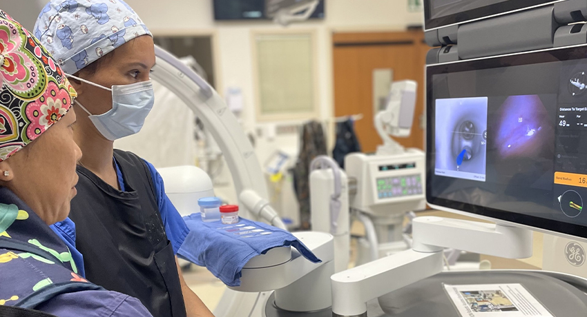
Nishi also utilizes AI in the surgical suite.
“Robotic bronchoscopy is an AI-enabled tool that allows us to navigate virtually to any area of the lung,” she said. “We can biopsy lesions with a high degree of accuracy and a much better safety profile than some of the other methods used to sample lung nodules.”
Nishi has seen significant progress during her career as a pulmonologist.
“When I trained, lung cancer was almost always a death sentence,” she said. “The treatment was as bad as the disease and people lived on average maybe five years. There were some new medicines, including immunotherapy and targeted therapies, coming out for lung cancer. Nobody knew what that would mean, but it was promising.”
Now, Nishi has seen many of those promising advances become clinical realities and believes AI also has an important role in the present and future of lung cancer diagnosis and care. Optimal screening remains an important part of improving patient outcomes.