


The Cardiac Cycle
|
|
|
||||||||
|
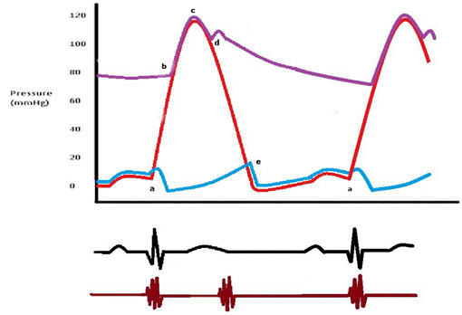
Figure 1. Cardiac cycle of the left side of the heart. The EKG below the diagram shows the corresponding waves with each phase of the cardiac cycle. The bottom line represents the first and second heart sounds. |
|||||||||
The cardiac cycle represents the hemodynamic and electric changes that occur in systole and diastole. It has many phases.
Phases of the Cardiac Cycle:
- Isovolumetric ventricular contraction (a-b): This phase marks the beginning of systole and starts with the appearance of the QRS complex on the EKG and the closure of the AV valves at point (a). With all valves closed, the ventricle generates positive pressure without any change in its volume (isovolumetric) to overcome the semilunar valves resistance that open at point b. This phase usually lasts for 6% of the cardiac cycle.
- Rapid ejection (b-c): As the semilunar valves open at point (b), there is a rapid ejection of blood due to increased ventricular contractility. The arterial pressure increases until reaching it maximum at point (c). This phase usually lasts for 13% of the cardiac cycle.
- Reduced ejection (c-d): This phase marks the beginning of ventricular repolarization as depicted by the onset of the T wave on the EKG. Repolarization leads to a rapid decline in ventricular pressures and hence the reduced rate of ejection. However, some forward flow of blood continues secondary to remnant kinetic energy from the previous phase. This phase usually lasts for 15% of the cardiac cycle.
- Isovolumetric relaxation (d-e): When the ventricular pressures drop below the diastolic aortic and pulmonary pressures (80 mmHg and 10 mmHg respectively), the aortic and pulmonary valves close producing the second heart sound (point d). This marks the beginning of diastole. The ventricles generate negative pressure without changing their volume (isovolumetric) so that the ventricular pressure becomes lower than the atrial pressure. This phase usually lasts for 8% of the cardiac cycle.
- Ventricular filling (e-a): As the AV valves open at point (e), ventricular filling starts. The initial rapid filling is mainly augmented by ventricular suction which results from ventricular untwisting and the return of each ventricular muscle fiber to its slack length. The ventricular pressure gradually increases until it equals the atrial pressure and the AV valves close (point a). This phase usually lasts for 44% of the cardiac cycle.
- Atrial contraction: Finally, near the end of ventricular diastole, the atrial contraction contributes about 10% of the ventricular filling volume. This is represented by the P wave on the EKG of the following cycle. This phase usually lasts for 14% of the cardiac cycle.
